. 2000 Dec;20(23):8923-32.
doi: 10.1128/MCB.20.23.8923-8932.2000.

Constitutive instability of muscle regulatory factor Myf5 is distinct from its mitosis-specific disappearance, which requires a D-box-like motif overlapping the basic domain

Affiliations

Constitutive instability of muscle regulatory factor Myf5 is distinct from its mitosis-specific disappearance, which requires a D-box-like motif overlapping the basic domain

C Lindon et al. Mol Cell Biol. 2000 Dec.

Abstract

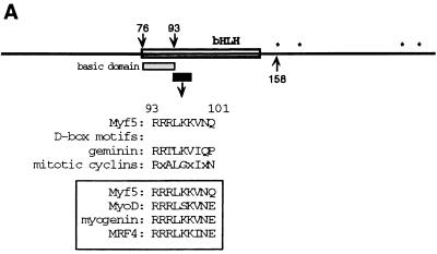
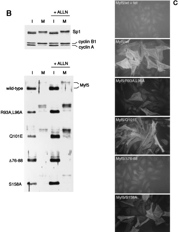
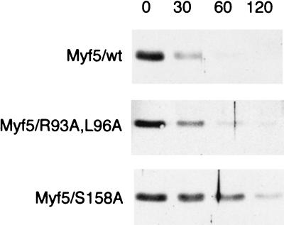
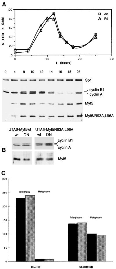
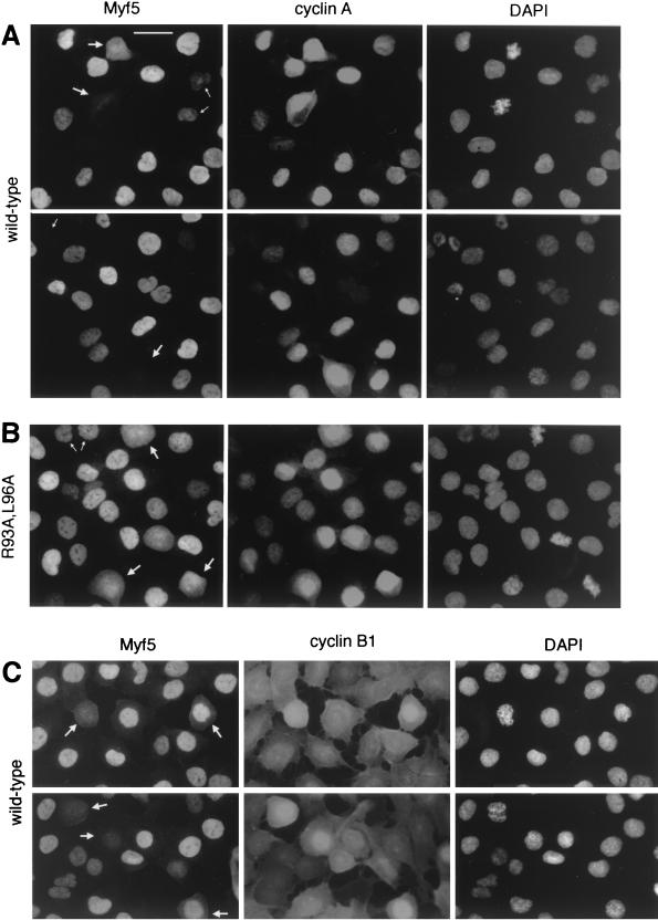
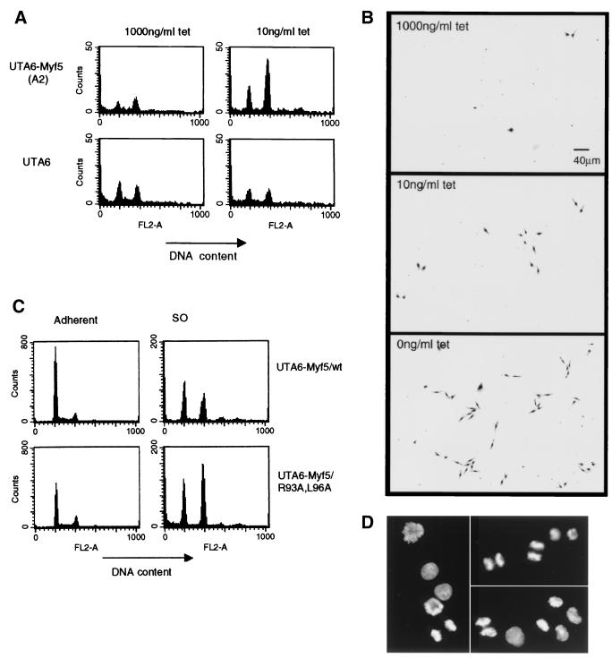
Transcription factors Myf5 and MyoD play critical roles in controlling myoblast identity and differentiation. In the myogenic cell line C2, we have found that Myf5 expression, unlike that of MyoD, is restricted to cycling cells and regulated by proteolysis at mitosis. In the present study, we have examined Myf5 proteolysis through stable transfection of myogenically convertible U20S cells with Myf5 derivatives under the control of a tetracycline-sensitive promoter. A motif within the basic helix-loop-helix domain of Myf5 (R93 to Q101) resembles the "destruction box" characteristic of substrates of mitotic proteolysis and thought to be recognized by the anaphase-promoting complex or cyclosome (APC). Mutation of this motif in Myf5 stabilizes the protein at mitosis but does not affect its constitutive turnover. Conversely, mutation of a serine residue (S158) stabilizes Myf5 in nonsynchronized cultures but not at mitosis. Thus, at least two proteolytic pathways control Myf5 levels in cycling cells. The mitotic proteolysis of Myf5 is unlike that which has been described for other destruction box-dependent substrates: down-regulation of Myf5 at mitosis appears to precede that of known targets of the APC and is not affected by a dominant-negative version of the ubiquitin carrier protein UbcH10, implicated in the APC-mediated pathway. Finally, we find that induction of Myf5 perturbs the passage of cells through mitosis, suggesting that regulation of Myf5 levels at mitosis may influence cell cycle progression of Myf5-expressing muscle precursor cells.


Figures

FIG. 1
FIG. 1
Stabilization of Myf5 at mitosis. (A) Schematic representation of Myf5 showing location of the D-box-like motif. The RXXL motif is present in all members of the MRF family (boxed) and resembles more closely the functional D-box in geminin than those in the mitotic cyclins. ∗, proline-directed serine/threonine residues that are possible phosphorylation sites at mitosis. (B) UTA6-derived cell lines were induced for expression of different versions of Myf5 by withdrawal of tetracycline from the culture medium for 16 h in the presence of nocodazole (to enrich the population of cells in mitosis). Parallel cultures were treated for 2 h with the proteasome inhibitor ALLN prior to preparation of extracts. Immunoblots of protein extracts prepared from the mitotic cells harvested by mitotic shake-off (M) and extracts prepared from the adherent, interphase (I) cells are compared for expression of Myf5. Levels of mitotic cyclins, and levels of transcription factor Sp1 as a loading control, are shown for a representative set of extracts (top). We note that steady-state levels of Myf5 proteins varied between clones, and we selected clones between which levels could most easily be compared. (C) UTA6-derived cell lines were induced for expression of different versions of Myf5 for 5 days. Cells were fixed and stained for expression of a skeletal muscle-specific marker, Troponin T, as described in Materials and Methods. All antibodies are described in Materials and Methods. tet, tetracycline.
FIG. 1
FIG. 1
Stabilization of Myf5 at mitosis. (A) Schematic representation of Myf5 showing location of the D-box-like motif. The RXXL motif is present in all members of the MRF family (boxed) and resembles more closely the functional D-box in geminin than those in the mitotic cyclins. ∗, proline-directed serine/threonine residues that are possible phosphorylation sites at mitosis. (B) UTA6-derived cell lines were induced for expression of different versions of Myf5 by withdrawal of tetracycline from the culture medium for 16 h in the presence of nocodazole (to enrich the population of cells in mitosis). Parallel cultures were treated for 2 h with the proteasome inhibitor ALLN prior to preparation of extracts. Immunoblots of protein extracts prepared from the mitotic cells harvested by mitotic shake-off (M) and extracts prepared from the adherent, interphase (I) cells are compared for expression of Myf5. Levels of mitotic cyclins, and levels of transcription factor Sp1 as a loading control, are shown for a representative set of extracts (top). We note that steady-state levels of Myf5 proteins varied between clones, and we selected clones between which levels could most easily be compared. (C) UTA6-derived cell lines were induced for expression of different versions of Myf5 for 5 days. Cells were fixed and stained for expression of a skeletal muscle-specific marker, Troponin T, as described in Materials and Methods. All antibodies are described in Materials and Methods. tet, tetracycline.
FIG. 2
FIG. 2
Stabilization of Myf5 in nonsynchronized cells. Cells were induced for 24 h and then CHX was added to cultures to block further protein synthesis. Protein extracts were prepared at the times indicated (in minutes following addition of CHX) and examined for Myf5 levels by immunoblot analysis.
FIG. 3
FIG. 3
Myf5 proteolysis at mitosis is distinct from that of mitotic cyclins. (A) UTA6-Myf5/ cells were synchronized at the start of S phase by a double thymidine block protocol (Materials and Methods). Cells were simultaneously released from the second thymidine block and induced for expression of Myf5 or Myf5/R93A,L96A. Samples were prepared for flow cytometric analysis of DNA content (top) and immunoblot analyses (bottom) at the times indicated. (B) UTA6-Myf5/wt and UTA6-Myf5/R93A,L96A cells were transfected with expression vectors for wild-type UbcH10 (lanes wt) or a dominant-negative version, UbcH10-DN (lanes DN). Total cell extracts were prepared after 48 h and examined by immunoblot analysis for levels of Myf5 proteins and mitotic cyclins. Transfection efficiencies were assessed by cotransfection with a β-galactosidase-expressing plasmid and were the same for each transfection (approximately 20%), as assessed by in situ staining for β-galactosidase activity in parallel cultures. (C) The transfected cell populations described in the Fig. 3B legend and identified by in situ staining for β-galactosidase activity were examined for the number of mitotic versus interphase figures. At least 250 transfected cells in 10 different fields were scored for each transfection. Black histograms, UTA6-Myf5/wt cells. Gray histograms, UTA6-Myf5/R93A,L96A cells.
FIG. 4
FIG. 4
Cell-by-cell analyses of Myf5 levels at mitosis. UTA6-Myf5/ cells were prepared as described for Fig. 3A and, 12 h after release from double thymidine block, were fixed and prepared for immunofluorescence analyses of Myf5 and cyclin A (A and B) or Myf5 and cyclin B1 (C). Bar, 25 μm. (A) UTA6-Myf5/wt cells costained for Myf5, cyclin A, and DAPI. Two fields are shown. Most cells show strong nuclear staining for cyclin A, confirming that the majority of cells are in late S phase, G2, or prophase. The redistribution of cyclin A throughout the cell is observed following nuclear-envelope breakdown at the end of prophase, and its levels decrease gradually during subsequent prometaphase and metaphase (31). Cells with decondensed chromatin and which show no cyclin A staining are presumed to be in early G1 (having completed mitosis), and indeed, these cells systematically occur in pairs (top, small arrows). As expected, there is a clear correlation between the intensities of Myf5 and cyclin A stainings in cells outside of mitosis. That is, Myf5 staining is strongest in cells showing strong nuclear cyclin A staining (but before the onset of nuclear condensation). Cyclin A-negative cells in late mitosis or G1 (bottom and top, respectively; small arrows) typically show faint or no staining for Myf5. By contrast, in prophase and prometaphase cells (large arrows), there is no clear correlation between Myf5 and cyclin A stainings. (B) UTA6-Myf5/R93A,L96A cells costained for Myf5, cyclin A, and DAPI. Small arrows, G1 cells; large arrows, prometaphase cells. (C) UTA6-Myf5/wt cells costained for Myf5, cyclin B1, and DAPI. Cyclin B1 translocates to the nucleus from the cytoplasm during prophase (31). Cells clearly in late prophase or prometaphase (showing nuclear cyclin B1, or in which nuclear-envelope breakdown has occurred) are indicated with arrows.
FIG. 5
FIG. 5
Flow cytometric analyses of Myf5-expressing cells. (A) Parental UTA6 cells or UTA6-Myf5/wt cells were cultured for 3 days at 1,000 or 10 ng of tetracycline (tet) per ml. The total cell population did not vary at these different doses of tetracycline. The culture medium was recovered after gentle shaking of the dish. Detached cells were harvested by centrifugation and prepared for flow cytometric analysis of DNA content. Samples were analyzed with a fixed time parameter to enable comparison of cell numbers in each sample. FL2-A (intensity of propidium iodide fluorescence) indicates relative DNA contents of cells, with a 4N cell population identified by an FL2-A of 400. Since the population of cells harvested resembles mitotic cells by phase-contrast microscopy, we suggest that the 2N fraction seen by flow cytometric analysis consists of cells which complete mitosis during the time of preparation of samples. (B) Detached cell populations harvested from UTA6-Myf5/ cells cultured at the doses of tetracycline indicated for 3 days were replated by transfer of the culture medium to fresh dishes. These cells were cultured for a further 24 h without change of medium and then fixed with 70% ethanol and stained with Giemsa (GIBCO-BRL). (C) Detached cell populations (SO cells) were prepared from UTA6-Myf/wt and UTA6/R93A,L96A cells grown for 3 days with 0 ng of tetracycline per ml. The adherent population of cells from each dish was recovered by trypsinization and prepared for flow cytometric analysis to allow quantification of the total cell population. Histograms from adherent and SO cells prepared with 0 ng of tetracycline per ml are shown (N.B., the adherent samples are diluted 50× compared to the SO samples). (D) Cells from the UTA6-Myf5/R93A,L96A SO population analyzed for panel C, fixed and stained with DAPI. Representative fields show cells in metaphase (left) and anaphase/telophase (top right) and undergoing cytokinesis (lower right).

Similar articles

Cited by

References

    1. Abu Hatoum O, Gross-Mesilaty S, Breitschopf K, Hoffman A, Gonen H, Ciechanover A, Bengal E. Degradation of myogenic transcription factor MyoD by the ubiquitin pathway in vivo and in vitro: regulation by specific DNA binding. Mol Cell Biol. 1998;18:5670–5677. - PMC - PubMed
    1. Albagli O, Lantoine D, Quief S, Quignon F, Kerckaert J, Montarras D, Pinset C, Lindon C. Overexpressed BCL6 (LAZ3) oncoprotein triggers apoptosis, delays S phase progression and associates with replication foci. Oncogene. 1999;18:5063–5075. - PubMed
    1. Arnold H-H, Winter B. Muscle differentiation: more complexity to the network of myogenic regulators. Curr Opin Genet Dev. 1998;8:539–544. - PubMed
    1. Auradé F, Pinset C, Chafey P, Gros F, Montarras D. Myf5, MyoD, myogenin and MRF4 myogenic derivatives of the embryonic mesenchymal cell line C3H10T1/2 exhibit the same adult muscle phenotype. Differentiation. 1994;55:185–192. - PubMed
    1. Bastians H, Topper L M, Gorbsky G, Ruderman J V. Cell cycle-regulated proteolysis of mitotic target proteins. Mol Biol Cell. 1999;10:3927–3941. - PMC - PubMed

Publication types