Comparative Study
. 2000 Dec;20(23):8996-9008.
doi: 10.1128/MCB.20.23.8996-9008.2000.

TAP (NXF1) belongs to a multigene family of putative RNA export factors with a conserved modular architecture

Affiliations
Comparative Study

TAP (NXF1) belongs to a multigene family of putative RNA export factors with a conserved modular architecture

A Herold et al. Mol Cell Biol. 2000 Dec.

Abstract

Vertebrate TAP (also called NXF1) and its yeast orthologue, Mex67p, have been implicated in the export of mRNAs from the nucleus. The TAP protein includes a noncanonical RNP-type RNA binding domain, four leucine-rich repeats, an NTF2-like domain that allows heterodimerization with p15 (also called NXT1), and a ubiquitin-associated domain that mediates the interaction with nucleoporins. Here we show that TAP belongs to an evolutionarily conserved family of proteins that has more than one member in higher eukaryotes. Not only the overall domain organization but also residues important for p15 and nucleoporin interaction are conserved in most family members. We characterize two of four human TAP homologues and show that one of them, NXF2, binds RNA, localizes to the nuclear envelope, and exhibits RNA export activity. NXF3, which does not bind RNA or localize to the nuclear rim, has no RNA export activity. Database searches revealed that although only one p15 (nxt) gene is present in the Drosophila melanogaster and Caenorhabditis elegans genomes, there is at least one additional p15 homologue (p15-2 [also called NXT2]) encoded by the human genome. Both human p15 homologues bind TAP, NXF2, and NXF3. Together, our results indicate that the TAP-p15 mRNA export pathway has diversified in higher eukaryotes compared to yeast, perhaps reflecting a greater substrate complexity.


Figures
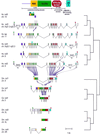
FIG. 1
FIG. 1
Intron-exon structure of the nxf genes. The domain organization of human TAP is indicated at the top. Hs, H. sapiens; Dm, D. melanogaster; Ce, C. elegans. When known, the chromosomal (chr.) locations are indicated below the gene names. Exons are colored according to the domains: purple, N-terminal portion found only in human homologues and C. elegans nxf1; yellow, RBD; green, LRR; red, NTF2-like domain; pink, linkers upstream and downstream of NTF2-like domain; cyan, UBA domain. 5′ and 3′ untranslated regions present in the ESTs or cDNA sequences are indicated by open boxes. Exons having no similarity to human TAP are gray. Introns are depicted as lines. The intron-exon structures are drawn to scale except for the long introns, which have breaks in the middle with the lengths indicated by numbers. An alternative splicing pathway is shown by lines above the gene (nxf2). A skipped exon is shown in black and connected by dotted lines (nxf3). Some characteristics of the sequences are indicated above the exon by filled triangles: green, initiation codon; red, termination codon; black, in-frame stop codon; cyan, frameshift. In nxf2, a stop codon created by alternative splicing is indicated as a red open triangle. On the complementary strand of nxf6, there is a region showing weak similarity to other TAP gene sequences; this region is shown in shift vertically. Some introns for the human TAP and NXF5 genes are not shown because the genes are mapped on distinct fragments of the genome sequences and the lengths of the introns are not clear. On the right, a simplified phylogenetic tree (not to scale) is shown.
FIG. 2
FIG. 2
Phylogenetic tree of NXF family sequences. The tree was drawn by the neighbor-joining method (34). Abbreviations (other than those in Fig. 1): Mm, Mus musculus; Rn, Rattus norvegicus; Sc, S. cerevisiae; Sp, S. pombe.
FIG. 3
FIG. 3
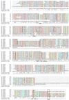
Multiple sequence alignment of NXF family sequences. First column, species names (Hs, H. sapiens; Dm, D. melanogaster; Ce, C. elegans); second column, protein names; third column, positions of the first aligned residues in each of the sequences. The positions conserved in 80% of the sequences are indicated in the consensus line: a, aromatic (FHWY); c, charged (DEHKR); h, hydrophobic (ACFGHIKLMRTVWY); l, aliphatic (LIV); o, hydroxyl (ST); p, polar (CDEHKNQRST); s, small (ACDGNPSTV); t, turnlike (ACDEGHKNQRST); u, tiny (AGS). The assigned domains are indicated below the consensus line. Highly conserved residues are indicated by colored boldface characters: orange, polar; light green, tiny; dark green, hydrophobic; blue, proline; light blue, hydroxyl; purple, cysteine. Exon boundaries are indicated by red marks.
FIG. 4
FIG. 4
Characterization of the N-terminal domains of NXF2 and NXF3. (A) GST pull-down assays were performed with [35S]methionine-labeled TAP, NXF2, NXF3, and the recombinant proteins indicated above the lanes. Lanes 2, background obtained with glutathione agarose beads coated with GST; lanes 3, proteins selected on immobilized GST-E1B-AP5 (fragment 101–453); lanes 4 to 6, binding to GST-REF1-II or fragments of this protein as indicated. In all panels, 1/10 of the inputs (lanes 1) and 1/3 of the bound fractions (lanes 2 to 6) were analyzed on SDS-PAGE followed by fluorography. Supernatant fractions were analyzed in parallel in order to confirm that the absence of binding was not due to protein degradation (data not shown). (B and C) Gel mobility retardation assays were performed using a radiolabeled RNA probe derived from pBS polylinker and purified recombinant proteins fused to GST. (B) TAP or NXF2 N-terminal fragments (50 ng) were used; (C), 1.5 μg of the corresponding RNA binding domains were used. Unlabeled competitor tRNA was added as indicated above the lanes. The position of the free RNA probe is indicated. The asterisk indicates the positions of the RNA-protein complexes. (D) A gel mobility retardation assay was performed with reticulocyte lysates unprogrammed (R) or programmed with cDNAs encoding TAP, NXF2, or NXF3. In lane 4, unlabeled M36 competitor RNA was added, while in lanes 5 and 6, CTE competitor RNA was included in the reaction mixtures. The amounts of the competitor RNAs are indicated above the lanes. The position of the free RNA probe is indicated on the left. (E) Schematic representation of pDM138 and pDM138-CTE vectors (14). (F) Quail cells were transfected with plasmids pDM138 or pDM138-CTE along with various plasmids encoding either GFP alone or fused to the N termini of TAP, NXF2, NXF3, or the TAP mutants indicated on the left. TAPΔNPC has a deletion of residues 541 to 613. The cells were collected 40 h after transfection, and CAT activity was determined. Data from three separate experiments were expressed relative to the activities measured when GFP alone was coexpressed with pDM138 with or without CTE. The data are means ± standard deviations.
FIG. 5
FIG. 5
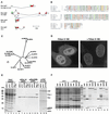
p15-2a, a human p15-1 homologue, interacts with TAP and localizes to the nuclear rim. (A) Intron-exon structures of p15 family sequences. Protein coding regions and untranslated regions are colored red and white, respectively. The positions of initiation and termination codons are indicated by green and red triangles, respectively. For human p15-2a and -b, the alternative splicing pathway is shown by lines above the gene. The 5′ exon of the p15-2a gene contains an open reading frame of five amino acids, while p15-2b gene 5′ exon sequences contain an in-frame stop codon but no in-frame ATG. Therefore, translation of this mRNA may generate a truncated protein starting at methionine 29 in the p15-2a sequence. Alternatively, translation may start at the GTG codon (green open triangle), resulting in an open reading frame (gray), since this region does not show any similarity to the other p15 sequences. (B) Multiple sequence alignment of p15 family sequences. The symbols are as in Fig. 3. (C) Phylogenetic tree of the NTF2 family sequences. The tree was drawn by the neighbor-joining method (34). Abbreviations (other than those in Fig. 1 and 2): At, Arabidopsis thaliana; Nt, Nicotiana tabacum; G3BP, Ras-GAP SH3 domain binding protein; MKK3, MAP kinase kinase 3; NPK2, a tobacco protein kinase. (D) Subcellular localization of p15-2a. HeLa cells were transfected with a pEGFP-N3 plasmid derivative expressing a zz fusion of p15-2a. The fusion protein was detected throughout the nucleoplasm and cytoplasm and was excluded from the nucleolus (left). On the right, HeLa cells were extracted with (+) Triton X-100 prior to fixation. A punctate labeling pattern was visible at the nuclear periphery for the p15-2a protein. (E) Lysates from E. coli expressing the Ran binding domain of Importin β (fragment 1–452) or supplemented with equimolar amounts of NTF2, p15-1, or p15-2a were incubated with IgG-Sepharose beads coated with purified zzRanGDP, zzRanQ69L-GTP, or zzTAP (fragment 61–619). After extensive washes, the bound proteins were eluted. One-hundredth of the inputs (lanes 1 to 4) and 1/10 of the bound fractions (lanes 6 to 16) were analyzed by SDS-PAGE followed by Coomassie blue staining. (F) Lysates from E. coli expressing GST fusions of TAP, NXF2, or NXF3 together with untagged versions of p15-1 or p15-2a were incubated with glutathione agarose beads. After extensive washes, the bound proteins were eluted with SDS sample buffer and analyzed by SDS-PAGE followed by Coomassie blue staining. Lanes 1 to 9, input lysates; lanes 11 to 19, bound fractions; lane 10, molecular mass markers (116, 97, 84, 66, 55, 45, 36, 29, 24, 20, and 14.2 kDa).
FIG. 6
FIG. 6
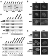
A fraction of NXF2 localizes to the nuclear rim. (A) GST pull-down assays were performed with [35S]methionine-labeled TAP, NXF2, or NXF3, and the recombinant proteins indicated above the lanes. One-tenth of the inputs (lanes 1, 4, and 7) and one-third of the bound fractions (lanes 2, 3, 5, 6, 8, and 9) were analyzed by SDS-PAGE followed by fluorography. (B) NXF2 interacts with multiple components of the NPC. GST pull-down assays were performed with the [35S]methionine-labeled nucleoporins indicated on the left of the panels and recombinant GST or TAP, TAP W594A, TAP D595R, or NXF2 fused to GST, as indicated above the lanes. One-tenth of the input (lane 1) and one-third of the bound fractions (lanes 2 to 6) were analyzed on SDS-PAGE followed by fluorography. (C) GST pull-down assays were performed with the [35S]methionine-labeled nucleoporins indicated on the left of the panels and recombinant GST, GST-NXF2, or various NXF2 mutants as indicated above the lanes. Samples were analyzed as indicated for panel A. (D and E) HeLa cells were transfected with pEGFP-C1 plasmid derivatives expressing GFP fusions of TAP, NXF2, and NFX3 or various TAP or NXF2 mutants as indicated on the left. Approximately 20 h after transfection, the cells were fixed in formaldehyde, permeabilized with Triton X-100, and directly observed with a fluorescence microscope. For all proteins, the GFP signal was detected throughout the nucleoplasm (−Tx). Cytoplasmic staining was also detected for NXF3. On the right (+Tx), HeLa cells were extracted with Triton X-100 prior to fixation. A punctate labeling pattern was visible at the nuclear periphery for TAP and NXF2, while in cells transfected with NXF3, TAP D595R, or the NXF2 mutants, no GFP signal was detected at the nuclear rim.
FIG. 7
FIG. 7
NXF2 exhibits general RNA nuclear export activity. (A to D) Human 293 cells were transfected with a mixture of plasmids encoding β-Gal, CAT, and either GFP alone (−) or fused to the N termini of TAP, NXF2, NXF3, and various TAP and NXF2 mutants as indicated on the left. pEGFP-N3 derivatives encoding zz-tagged versions of p15-1 and p15-2a were cotransfected as indicated. The cells were collected 40 h after transfection, and β-Gal and CAT activities were determined. Data from three separate experiments were expressed relative to the activities measured when GFP alone was coexpressed with pDM138. The data are means ± standard deviations. (B and C) Protein expression levels were analyzed by Western blotting with anti-GFP antibodies. (B) Arrowheads indicate the positions of NXF proteins fused to GFP or of GFP itself, while the asterisks show the positions of p15 proteins.

References

    1. Adams M D, Celniker S E, Holt R A, Evans C A, Gocayne J D, Amanatides P G, Scherer S E, Li P W, Hoskins R A, et al. The genome sequence of Drosophila melanogaster. Science. 2000;287:2185–2195. - PubMed
    1. Almeida F, Saffrich R, Ansorge W, Carmo-Fonseca M. Microinjection of anti-coilin antibodies affects the structure of coiled bodies. J Cell Biol. 1998;142:899–912. - PMC - PubMed
    1. Altschul S F, Madden T L, Schaffer A A, Zhang J, Zhang Z, Miller W, Lipman D J. Gapped BLAST and PSI-BLAST: a new generation of protein database search programs. Nucleic Acids Res. 1997;25:3389–3402. - PMC - PubMed
    1. Bachi A, Braun I C, Rodrigues J P, Panté N, Ribbeck K, von Kobbe C, Kutay U, Wilm M, Görlich D, Carmo-Fonseca M, Izaurralde E. The C-terminal domain of TAP interacts with the nuclear pore complex and promotes export of specific CTE-bearing RNA substrates. RNA. 2000;6:136–158. - PMC - PubMed
    1. Bear J, Tan W, Zolotukhin A S, Tabernero C, Hudson E A, Felber B K. Identification of novel import and export signals of human TAP, the protein that binds to the CTE element of the type D retrovirus mRNAs. Mol Cell Biol. 1999;19:6306–6317. - PMC - PubMed

Publication types

MeSH terms

Associated data