. 2000 Dec;20(23):9018-27.
doi: 10.1128/MCB.20.23.9018-9027.2000.

SU6656, a selective src family kinase inhibitor, used to probe growth factor signaling

Affiliations

SU6656, a selective src family kinase inhibitor, used to probe growth factor signaling

R A Blake et al. Mol Cell Biol. 2000 Dec.

Abstract

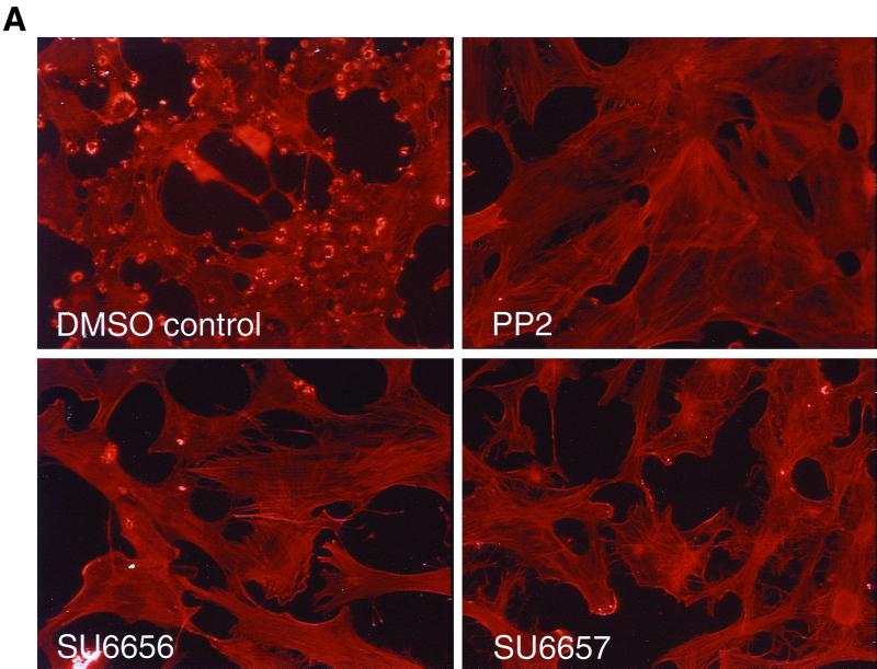
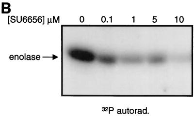
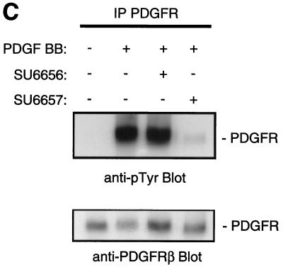
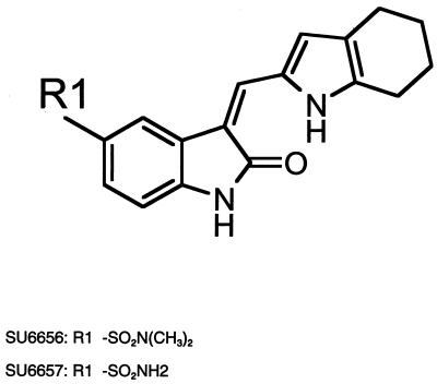
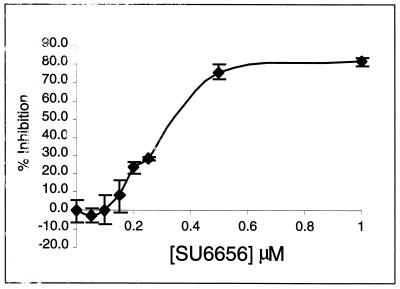
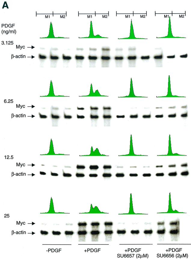
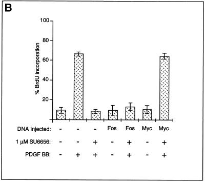
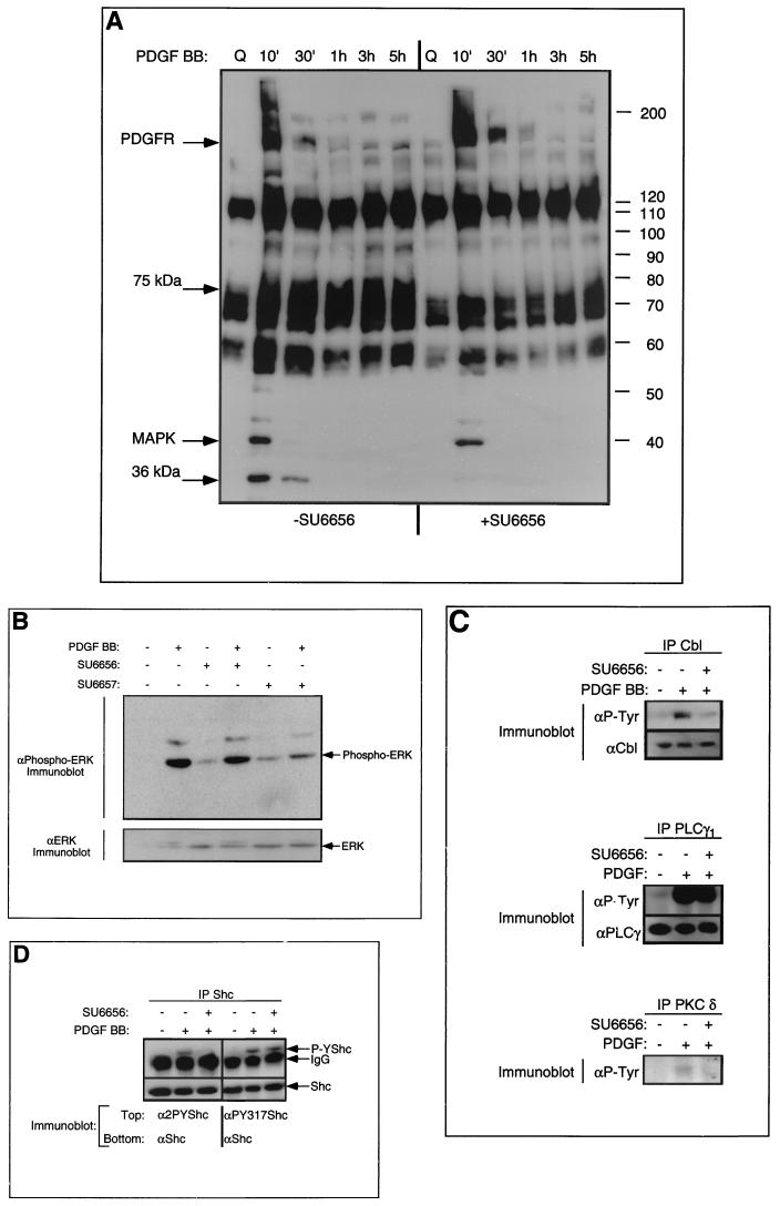
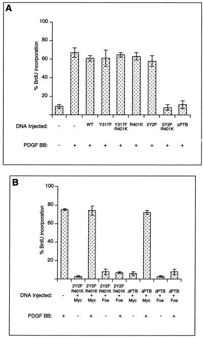
The use of small-molecule inhibitors to study molecular components of cellular signal transduction pathways provides a means of analysis complementary to currently used techniques, such as antisense, dominant-negative (interfering) mutants and constitutively activated mutants. We have identified and characterized a small-molecule inhibitor, SU6656, which exhibits selectivity for Src and other members of the Src family. A related inhibitor, SU6657, inhibits many kinases, including Src and the platelet-derived growth factor (PDGF) receptor. The use of SU6656 confirmed our previous findings that Src family kinases are required for both Myc induction and DNA synthesis in response to PDGF stimulation of NIH 3T3 fibroblasts. By comparing PDGF-stimulated tyrosine phosphorylation events in untreated and SU6656-treated cells, we found that some substrates (for example, c-Cbl, and protein kinase C delta) were Src family substrates whereas others (for example, phospholipase C-gamma) were not. One protein, the adaptor Shc, was a substrate for both Src family kinases (on tyrosines 239 and 240) and a distinct tyrosine kinase (on tyrosine 317, which is perhaps phosphorylated by the PDGF receptor itself). Microinjection experiments demonstrated that a Shc molecule carrying mutations of tyrosines 239 and 240, in conjunction with an SH2 domain mutation, interfered with PDGF-stimulated DNA synthesis. Deletion of the phosphotyrosine-binding domain also inhibited synthesis. These inhibitions were overcome by heterologous expression of Myc, supporting the hypothesis that Shc functions in the Src pathway. SU6656 should prove a useful additional tool for further dissecting the role of Src kinases in this and other signal transduction pathways.


Figures

FIG. 1
FIG. 1
Effects of PP2, SU6656, and SU6657. (A) NIH 3T3 cells overexpressing the activated mutant SrcY527F were treated for 16 h with the compound (5 μM) indicated in each panel. The cells were then fixed with 3% paraformaldehyde in phosphate-buffered saline and stained with Texas red-conjugated phalloidin. (B) Immune complex kinase assay of Src with enolase as a substrate. 32P-labeled enolase was visualized by autoradiography (32P/autorad.). (C) NIH 3T3 cells were treated (indicated by + in the corresponding row) with 5 μM SU6656, SU6657, or dimethyl sulfoxide control (final concentration, 0.1% [vol/vol]) for 1 h prior to stimulation of the cells with 25 ng of PDGF BB/ml for 10 min. The upper blot shows an anti-phosphotyrosine blot of PDGFRβ immunoprecipitated (IP) from each of the samples. Below is the same blot stripped and reprobed for the level of PDGFRβ protein.
FIG. 1
FIG. 1
Effects of PP2, SU6656, and SU6657. (A) NIH 3T3 cells overexpressing the activated mutant SrcY527F were treated for 16 h with the compound (5 μM) indicated in each panel. The cells were then fixed with 3% paraformaldehyde in phosphate-buffered saline and stained with Texas red-conjugated phalloidin. (B) Immune complex kinase assay of Src with enolase as a substrate. 32P-labeled enolase was visualized by autoradiography (32P/autorad.). (C) NIH 3T3 cells were treated (indicated by + in the corresponding row) with 5 μM SU6656, SU6657, or dimethyl sulfoxide control (final concentration, 0.1% [vol/vol]) for 1 h prior to stimulation of the cells with 25 ng of PDGF BB/ml for 10 min. The upper blot shows an anti-phosphotyrosine blot of PDGFRβ immunoprecipitated (IP) from each of the samples. Below is the same blot stripped and reprobed for the level of PDGFRβ protein.
FIG. 2
FIG. 2
Structures of SU6656 and SU6657.
FIG. 3
FIG. 3
Inhibition of PDGF-stimulated NIH 3T3 cell DNA synthesis. NIH 3T3 cells seeded on glass coverslips were made quiescent for 24 h in 0.5% FCS and treated with various concentrations of SU6656. After 2 h, the cells were stimulated with 25 ng of PDGF BB/ml in the presence of BrdU for 24 h and then fixed and stained for the incorporation of BrdU into newly synthesized DNA. The data is presented as percent inhibition (± standard deviation) of the PDGF BB-stimulated DNA synthesis. Of the PDGF BB-stimulated cells, 70.5% incorporated BrdU compared to 11.7% of nonstimulated cells.
FIG. 4
FIG. 4
SU6656 inhibits PDGF-stimulated c-Myc induction. (A) Effects of SU6656 on PDGF-stimulated c-Myc expression and DNA synthesis. Results of RPAs and FACS analyses performed with cells treated with various concentrations of PDGF BB in combination with SU6656 (2 μM) or SU6657 (2 μM) as indicated are presented. For RPAs, experiments were done in triplicate for each treatment condition. The c-Myc signals were quantitated on a PhosphorImager and corrected based on the actin signal, and then percent inhibition with the two inhibitors was calculated (Table 2). For FACS analyses, cells that did not and did contain newly synthesized DNA were gated as M1 and M2, respectively. Quantitation involved comparing the percentages of cells in M2 under various treatment conditions and is shown in Table 2. (B) Myc rescues SU6656 inhibition. NIH 3T3 cells were plated on coverslips and serum starved for 30 h. The cells were microinjected into the nucleus with plasmids encoding c-Myc or c-Fos along with a marker. Fourteen hours later, the cells were treated with 1 μM SU6656 or dimethyl sulfoxide for 1 h followed by PDGF BB stimulation (20 ng/ml) and BrdU labeling for 24 h. The cells were fixed and stained for marker and BrdU incorporation by indirect immunofluorescence and analyzed by microscopic examination. The data are presented as the percent (± standard deviation) BrdU-positive cells present in expressing and nonexpressing cells under the specified conditions. The data are representative of three independent experiments with 100 to 300 expressing cells in each case. +, present; −, absent.
FIG. 4
FIG. 4
SU6656 inhibits PDGF-stimulated c-Myc induction. (A) Effects of SU6656 on PDGF-stimulated c-Myc expression and DNA synthesis. Results of RPAs and FACS analyses performed with cells treated with various concentrations of PDGF BB in combination with SU6656 (2 μM) or SU6657 (2 μM) as indicated are presented. For RPAs, experiments were done in triplicate for each treatment condition. The c-Myc signals were quantitated on a PhosphorImager and corrected based on the actin signal, and then percent inhibition with the two inhibitors was calculated (Table 2). For FACS analyses, cells that did not and did contain newly synthesized DNA were gated as M1 and M2, respectively. Quantitation involved comparing the percentages of cells in M2 under various treatment conditions and is shown in Table 2. (B) Myc rescues SU6656 inhibition. NIH 3T3 cells were plated on coverslips and serum starved for 30 h. The cells were microinjected into the nucleus with plasmids encoding c-Myc or c-Fos along with a marker. Fourteen hours later, the cells were treated with 1 μM SU6656 or dimethyl sulfoxide for 1 h followed by PDGF BB stimulation (20 ng/ml) and BrdU labeling for 24 h. The cells were fixed and stained for marker and BrdU incorporation by indirect immunofluorescence and analyzed by microscopic examination. The data are presented as the percent (± standard deviation) BrdU-positive cells present in expressing and nonexpressing cells under the specified conditions. The data are representative of three independent experiments with 100 to 300 expressing cells in each case. +, present; −, absent.
FIG. 5
FIG. 5
Effect of SU6656 on tyrosine phosphorylation. (A) Serum-starved, quiescent NIH 3T3 cells were treated with SU6656 (2 μM) or mock dimethyl sulfoxide (DMSO) treated for 1 h followed by PDGF BB stimulation (20 ng/ml). Lysates were made at the indicated time points. Total cell lysate (25 μg) was resolved for each sample on SDS-polyacrylamide gel electrophoresis (PAGE), transferred to Immobilon, and probed with anti-phosphotyrosine antibodies followed by enhanced chemiluminescent detection. MAPK, MAP kinase. (B) NIH 3T3 cells were treated with (+) either SU6656 or SU6657 (2 μM) or DMSO alone (10 min) as indicated, then stimulated for 10 min with 25 ng of PDGF BB/ml. The cells were lysed in RIPA buffer, and the protein was resolved on SDS-PAGE followed by immunoblotting with a monoclonal antibody specific for tyrosine-phosphorylated ERK1 and -2 (upper blot). The lower blot shows immunoblotting for the level of ERK2. (C) Serum-starved, quiescent NIH 3T3 cells were treated with SU6656 (2 μM) or mock DMSO treated for 1 h followed by PDGF BB stimulation (20 ng/ml). Lysates were made at 10 min following stimulation, and the proteins indicated were immunoprecipitated. Samples were resolved on SDS-PAGE, transferred to Immobilon, and probed with anti-phosphotyrosine antibodies followed by enhanced chemiluminescent detection (upper blot in each case). The immunoblots were then stripped and reprobed for levels (lower blot in each case), except for PKC δ (data not shown). (D) Serum-starved, quiescent NIH 3T3 cells were treated with SU6656 (2 μM), or mock DMSO treated for 1 h followed by PDGF BB stimulation (20 ng/ml). Lysates were made at 10 min following stimulation, and Shc protein was immunoprecipitated. Samples were resolved on SDS-PAGE, transferred to Immobilon, and probed with either anti-2PYShc (anti-Shc pY239-pY240) (lanes 1 to 3) or anti-PY317Shc (lanes 4 to 6) followed by enhanced chemiluminescent detection (upper blot in each case). The immunoblots were then stripped and reprobed for Shc levels (lower blot in each case). IgG, immunoglobulin G.
FIG. 6
FIG. 6
Shc is part of the Src pathway. (A) NIH 3T3 cells were plated on coverslips and serum starved for 30 h. The cells were microinjected into the nucleus with plasmids encoding tagged Shc wild-type (WT) and mutant proteins. Fourteen hours later, the cells were stimulated with (+) PDGF BB (20 ng/ml) and BrdU labeled for 24 h. The cells were fixed and stained for Shc expression and BrdU incorporation by indirect immunofluorescence. The data are presented as the percent (± standard deviation [SD]) BrdU-positive cells present among expressing and nonexpressing cells under the specified conditions. The data are representative of three independent experiments with 100 to 300 expressing cells in each case. (B) NIH 3T3 cells were plated on coverslips and serum starved for 30 h. The cells were microinjected into the nucleus with mixtures of plasmids encoding c-Myc and HA-Shc mutants or c-Fos and HA-Shc mutants. Fourteen hours later, the cells were stimulated with PDGF BB (20 ng/ml) and BrdU labeled for 24 h. The cells were fixed and stained for HA-Shc expression and BrdU incorporation by indirect immunofluorescence. The data are presented as the percent (± SD) BrdU-positive cells present among expressing and nonexpressing cells under the specified conditions. The data are representative of three independent experiments with 100 to 300 expressing cells in each case.

References

    1. Barone M V, Courtneidge S A. Myc but not Fos rescue of PDGF signalling block caused by kinase inactive Src. Nature. 1995;378:509–512. - PubMed
    1. Blake R A, Garcia-Paramio P, Parker P J, Courtneidge S A. Src promotes PKCdelta degradation. Cell Growth Differ. 1999;10:231–241. - PubMed
    1. Broome M A, Courtneidge S A. No requirement for src family kinases for PDGF signaling in fibroblasts expressing SV40 large T antigen. Oncogene. 2000;19:2867–2869. - PubMed
    1. Broome M A, Galisteo M L, Schlessinger J, Courtneidge S A. The proto-oncogene c-Cbl is a negative regulator of DNA synthesis initiated by both receptor and cytoplasmic tyrosine kinases. Oncogene. 1999;18:2908–2912. - PubMed
    1. Cardenas M E, Sanfridson A, Cutler N S, Heitman J. Signal-transduction cascades as targets for therapeutic intervention by natural products. Trends Biotechnol. 1998;16:427–433. - PubMed

MeSH terms

LinkOut - more resources