Comparative Study
. 2000 Dec;20(23):9041-54.
doi: 10.1128/MCB.20.23.9041-9054.2000.

Transforming growth factor beta-independent shuttling of Smad4 between the cytoplasm and nucleus

Affiliations
Comparative Study

Transforming growth factor beta-independent shuttling of Smad4 between the cytoplasm and nucleus

C E Pierreux et al. Mol Cell Biol. 2000 Dec.

Abstract

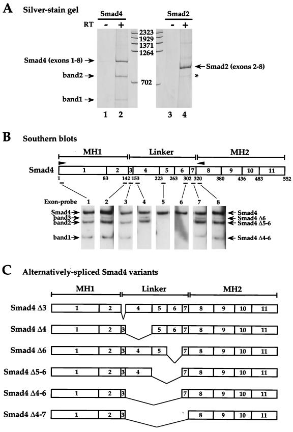
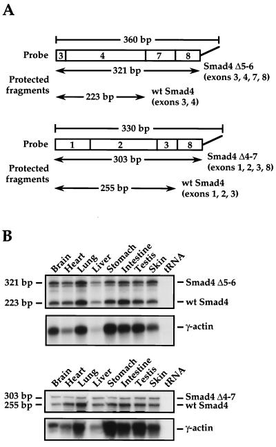
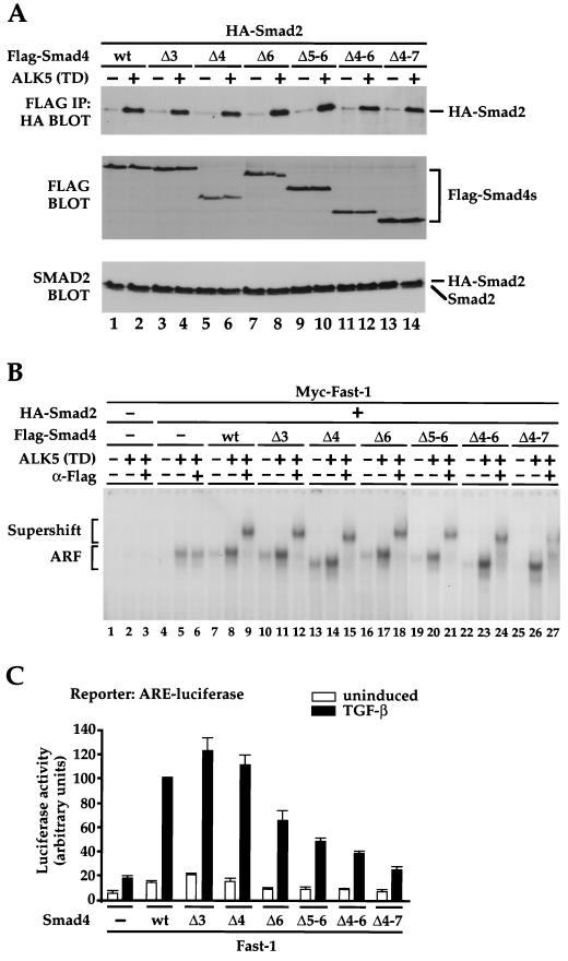
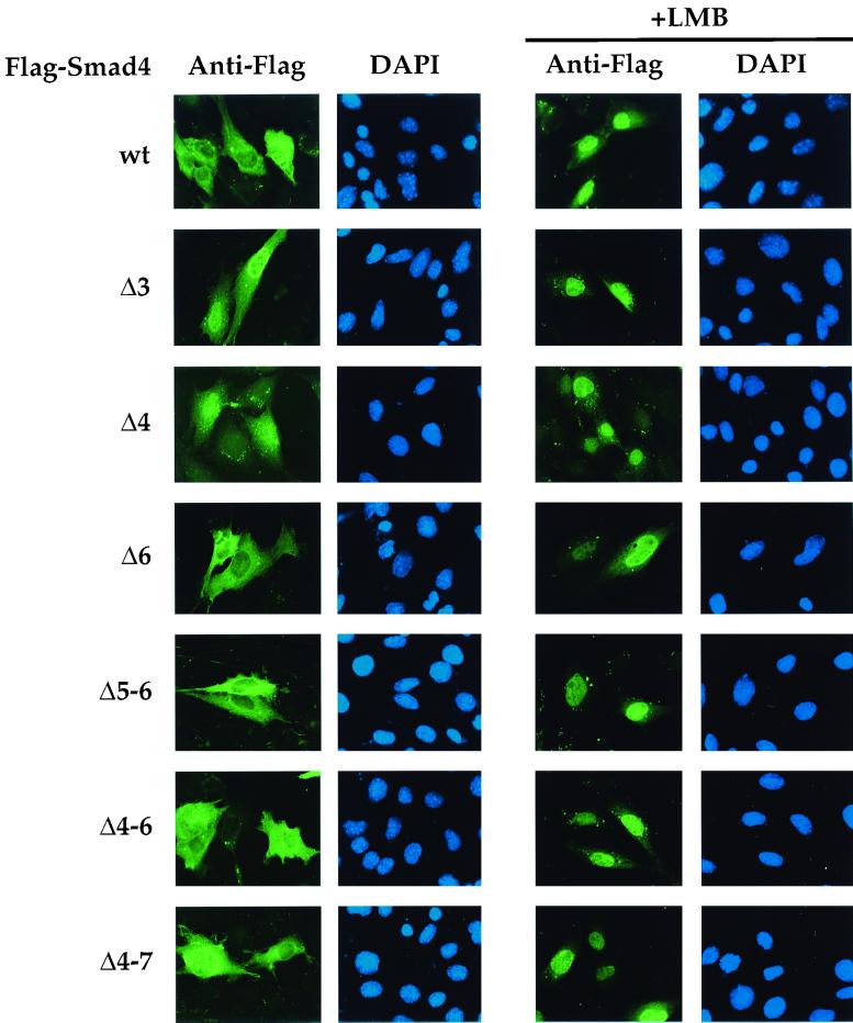
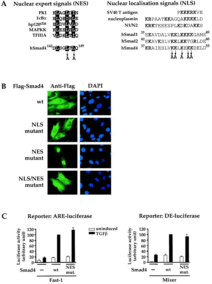
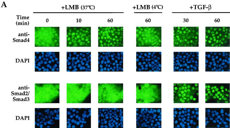
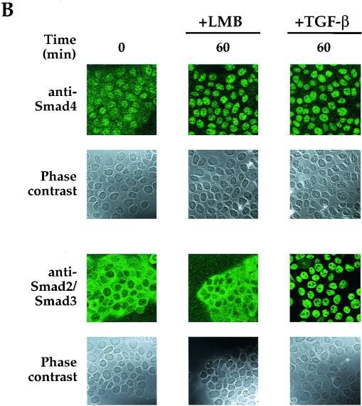
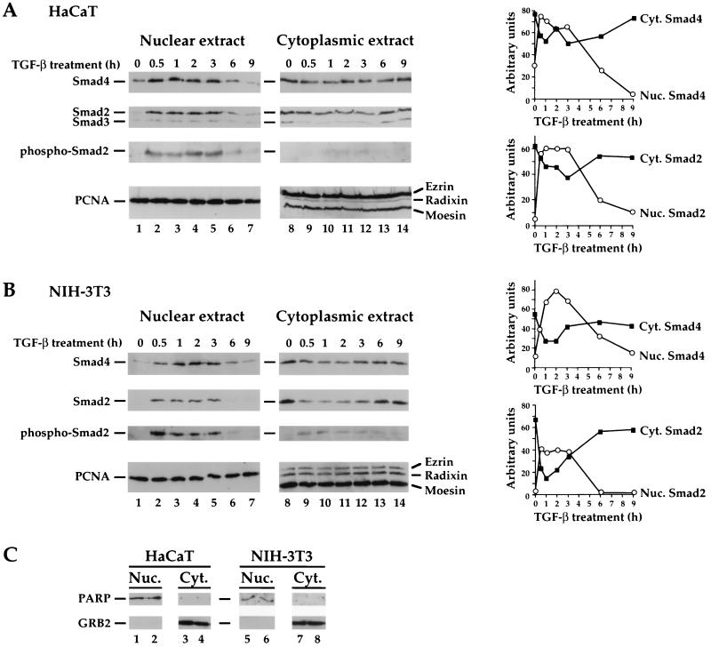
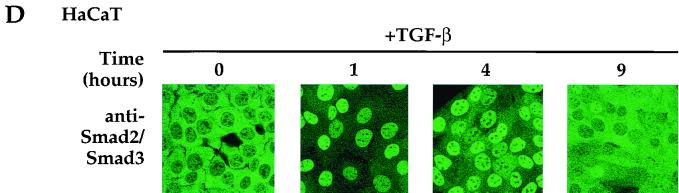
Smad4 plays a pivotal role in all transforming growth factor beta (TGF-beta) signaling pathways. Here we describe six widely expressed alternatively spliced variants of human Smad4 with deletions of different exons in the linker, the region of Smad4 that separates the two well-conserved MH1 and MH2 domains. All these Smad4 variants form complexes with activated Smad2 and Smad3 and are incorporated into DNA-binding complexes with the transcription factor Fast-1, regardless of the amount of linker they contain. However, sequences encoded by exons 5 to 7 in the linker are essential for transcriptional activation. Most importantly, our observation that different Smad4 isoforms have different subcellular localizations has led us to the identification of a functional CRM1-dependent nuclear export signal in the Smad4 linker and a constitutively active nuclear localization signal in the N-terminal MH1 domain. In the absence of TGF-beta signaling, we conclude that Smad4 is rapidly and continuously shuttling between the nucleus and the cytoplasm, the distribution of Smad4 between the nucleus and the cytoplasm being dictated by the relative strengths of the nuclear import and export signals. We demonstrate that inhibition of CRM1-mediated nuclear export by treatment of cells with leptomycin B results in endogenous Smad4 accumulating very rapidly in the nucleus. Endogenous Smad2 and Smad3 are completely unaffected by leptomycin B treatment, indicating that the nucleocytoplasmic shuttling is specific for Smad4. We propose that, upon TGF-beta signaling, complex formation between Smad4 and activated Smad2 or -3 leads to nuclear accumulation of Smad4 through inhibition of its nuclear export. We demonstrate that after prolonged TGF-beta signaling Smad2 becomes dephosphorylated and Smad2 and Smad4 accumulate back in the cytoplasm.


Figures

FIG. 1
FIG. 1
Human Smad4 mRNA is alternatively spliced. (A) Silver-stained polyacrylamide gel showing fragments obtained from RT-PCRs performed using total RNA from HaCaT cells as template and forward and reverse primers specific for exon 1 and exon 8, respectively, of human Smad4 (lanes 1 and 2) or exon 2 and exon 8, respectively, of human Smad2 (lanes 3 and 4). The reactions in lanes 1 and 3 were performed with no reverse transcriptase. DNA markers are measured in base pairs. The fragment derived from full-length Smad4 is designated Smad4 (exons 1-8), and that derived from full-length Smad2 is designated Smad2 (exons 2-8). Bands 1 and 2 are derived from alternatively spliced Smad4 mRNA. The asterisk denotes the Smad2 alternatively spliced variant (Smad2 Δexon 3) (49). (B) Identification of Smad4 isoforms by Southern blotting. A diagram of the Smad4 coding region is shown, denoting the MH1, linker, and MH2 domains. Arrowheads show PCR primers used in the RT-PCR. Exons are denoted as boxes with the exon numbers indicated. Numbers below are amino acids at the exon-intron boundaries (13). Positions of the oligonucleotide exon-specific probes are indicated. RT-PCR products similar to that in lane 2 above were Southern blotted with exon-specific probes as indicated. The bands 1 and 2 are as described above; band 3 is detected only by Southern blotting. The bands are identified as Smad4 Δ4-6, Δ5-6, and Δ6, respectively. (C) Schematics of the alternatively spliced Smad4 isoforms identified.
FIG. 2
FIG. 2
The alternatively spliced Smad4 variants are widely expressed in the adult mouse. (A) Diagram of the RNase protection probes designed to specifically protect Smad4 Δ5-6 mRNA and Smad4 Δ4-7 mRNA and the sizes of the resulting protected fragments. In both cases, the probes also protect full-length Smad4 mRNA, giving rise to a second, smaller fragment. (B) RNase protection assays detecting Smad4 Δ5-6 (upper panels) or Smad4 Δ4-7 (lower panels) in different adult mouse tissues. Protected fragments corresponding to the alternatively spliced mRNA variants and full-length Smad4 mRNA are indicated. γ-Actin was used as a loading control for all tissues. wt, wild type.
FIG. 3
FIG. 3
Sequences in the Smad4 linker are not required for formation of transcription factor complexes but are required for transcriptional activation. (A) The Smad4 linker is not required for interaction with activated Smad2. Extracts were prepared from NIH 3T3 cells transfected with the different Flag-tagged alternatively spliced Smad4 variants with HA-Smad2, with or without constitutively active ALK5 [ALK5 (TD)] as indicated. Extracts were assayed either by immunoprecipitation of complexes with anti-Flag antibody followed by Western blotting with anti-HA antibody (top panel) or by Western blotting the whole extract with anti-Flag antibody (middle panel) or with anti-Smad2 antibody (bottom panel). (B) The Smad4 linker is not required for formation of the Fast-1–Smad2–Smad4 complex ARF on the ARE. Extracts were prepared from NIH 3T3 cells transfected with Myc–Fast-1, HA-Smad2, and Flag-tagged alternatively spliced Smad4 variants and ALK5 (TD) as indicated. The extracts were analyzed by band shift using the ARE as probe in the presence or absence of anti-Flag antibody (α-Flag) as indicated to confirm the presence of the Flag-tagged Smad4 spliced variant in the complex. The Fast-1–Smad2–Smad4 complex ARF is indicated, as is the supershifted ARF complex. (C) Sequences encoded by exons 5, 6, and 7 are required for transcriptional activation mediated by Smad4. MDA-MB468 cells were transfected with the ARE-luciferase reporter, plasmids expressing Fast-1, and alternatively spliced Smad4 variants as indicated. Cells were cultured with or without TGF-β1 (2 ng/ml) for 6 h. Cells were harvested, and luciferase activity was measured relative to LacZ activity from the internal control. The data are averaged from at least four independent experiments and were normalized by setting the TGF-β induction mediated by wild-type Smad4 to 100%. IP, immunoprecipitation; wt, wild type.
FIG. 4
FIG. 4
Subcellular localization of the alternatively spliced Smad4 variants in the absence and presence of LMB. NIH 3T3 cells were transfected with Flag-tagged Smad4 isoforms as indicated. (Left) The subcellular localization of the Smad4 isoforms in untreated cells was determined by indirect immunofluorescence using the anti-Flag antibody, and nuclei of the same cells were also stained with DAPI as indicated. (Right) Subcellular localization of the Smad4 isoforms in cells treated with LMB at a 20-ng/ml final concentration for 1 h. wt, wild type.
FIG. 5
FIG. 5
Smad4 contains an NES and an NLS. (A) The putative NES encoded by exon 3 of Smad4 is aligned with other previously characterized NESs (21, 43). Leucines 146 and 148 were mutated to alanine to generate the NES mutant as shown. The putative NLS of Smad4 is aligned with well-characterized NLSs (7) and with the putative NLSs of the R-Smads, Smad1 and Smad2. Lysines 45, 46, 48, 50, and 51 were mutated to alanine to generate the Smad4 NLS mutant. (B) The NES and NLS of Smad4 are functional. NIH 3T3 cells were transfected with Flag-tagged Smad4 derivatives, and their subcellular localization was determined as described for Fig. 4. (C) Smad4 forms functional complexes with activated Smad2 and Fast-1 or Mixer in the nucleus. MDA-MB468 cells were transfected with the appropriate luciferase reporters, plasmids expressing transcription factors (Fast-1 or Mixer), and wild-type or mutant Smad4 as shown. TGF-β inductions and luciferase assays were performed as described for Fig. 3. The data are averaged from at least three independent experiments, which were normalized by setting the TGF-β induction mediated by wild-type Smad4 to 100%. PKI, cyclic AMP-dependent protein kinase inhibitor; MAPKK, mitogen-activated protein kinase kinase; wt, wild type; mut., mutant; SV40, simian virus 40.
FIG. 6
FIG. 6
Endogenous Smad4 rapidly shuttles between the nucleus and the cytoplasm in unstimulated cells. (A) HaCaT cells were treated with LMB (20 ng/ml) for the times shown at either 37 or 4°C or with 2 ng of TGF-β per ml and then processed for immunofluorescence using either an anti-Smad4 monoclonal antibody or an anti-Smad2 and -Smad3 monoclonal antibody. Nuclei of the same cells were also stained with DAPI. Fluorescence was visualized using a Zeiss Axioplan upright fluorescence microscope. (B) Fields from a subset of the samples shown in panel A were examined by confocal laser scanning microscopy using a Zeiss LSM 510 confocal microscope. Phase-contrast images of the same fields are also shown.
FIG. 6
FIG. 6
Endogenous Smad4 rapidly shuttles between the nucleus and the cytoplasm in unstimulated cells. (A) HaCaT cells were treated with LMB (20 ng/ml) for the times shown at either 37 or 4°C or with 2 ng of TGF-β per ml and then processed for immunofluorescence using either an anti-Smad4 monoclonal antibody or an anti-Smad2 and -Smad3 monoclonal antibody. Nuclei of the same cells were also stained with DAPI. Fluorescence was visualized using a Zeiss Axioplan upright fluorescence microscope. (B) Fields from a subset of the samples shown in panel A were examined by confocal laser scanning microscopy using a Zeiss LSM 510 confocal microscope. Phase-contrast images of the same fields are also shown.
FIG. 7
FIG. 7
Prolonged TGF-β signaling leads to export of Smad2 and Smad4 from the nucleus to the cytoplasm. (A and B) Nuclear and cytoplasmic extracts were made from HaCaT cells (A) or NIH 3T3 cells (B) that were treated with 20 μg of cycloheximide per ml and were either uninduced or induced with TGF-β (2 ng/ml) for the times shown. They were analyzed by Western blotting with antibodies against Smad4 or Smad2 and -3 (as in Fig. 6); phosphorylated Smad2; the ERM proteins ezrin, radixin, and moesin; or PCNA. Nuclear and cytoplasmic extracts were from the same cells. Cytoplasmic extracts were concentrated to the same volume as nuclear extracts, and equal volumes were loaded on the SDS-polyacrylamide gel. The Western blots were quantitated, and the results are presented graphically (right panels). (C) Western blotting of the nuclear and cytoplasmic extracts from two time points with antibodies against the cytoplasmic protein GRB2 and the nuclear protein poly(ADP-ribose) polymerase (PARP) indicates that the extracts are virtually free from cross-contamination. (D) TGF-β-dependent nuclear import of Smad2 and Smad3 and export after prolonged signaling in HaCaT cells as detected by immunofluorescence. Cells were treated with 20 μg of cycloheximide per ml and were either uninduced or induced with TGF-β (2 ng/ml) for 1, 4, or 9 h and processed for immunofluorescence as described for Fig. 6. Cells were examined by confocal laser scanning microscopy using a Zeiss LSM 510 confocal microscope. Nuc., nuclear; Cyt., cytoplasmic.
FIG. 7
FIG. 7
Prolonged TGF-β signaling leads to export of Smad2 and Smad4 from the nucleus to the cytoplasm. (A and B) Nuclear and cytoplasmic extracts were made from HaCaT cells (A) or NIH 3T3 cells (B) that were treated with 20 μg of cycloheximide per ml and were either uninduced or induced with TGF-β (2 ng/ml) for the times shown. They were analyzed by Western blotting with antibodies against Smad4 or Smad2 and -3 (as in Fig. 6); phosphorylated Smad2; the ERM proteins ezrin, radixin, and moesin; or PCNA. Nuclear and cytoplasmic extracts were from the same cells. Cytoplasmic extracts were concentrated to the same volume as nuclear extracts, and equal volumes were loaded on the SDS-polyacrylamide gel. The Western blots were quantitated, and the results are presented graphically (right panels). (C) Western blotting of the nuclear and cytoplasmic extracts from two time points with antibodies against the cytoplasmic protein GRB2 and the nuclear protein poly(ADP-ribose) polymerase (PARP) indicates that the extracts are virtually free from cross-contamination. (D) TGF-β-dependent nuclear import of Smad2 and Smad3 and export after prolonged signaling in HaCaT cells as detected by immunofluorescence. Cells were treated with 20 μg of cycloheximide per ml and were either uninduced or induced with TGF-β (2 ng/ml) for 1, 4, or 9 h and processed for immunofluorescence as described for Fig. 6. Cells were examined by confocal laser scanning microscopy using a Zeiss LSM 510 confocal microscope. Nuc., nuclear; Cyt., cytoplasmic.

Similar articles

Cited by

References

    1. Chen C H, von Kessler D P, Park W, Wang B, Ma Y, Beachy P A. Nuclear trafficking of Cubitus interruptus in the transcriptional regulation of Hedgehog target gene expression. Cell. 1999;98:305–316. - PubMed
    1. Chen X, Rubock M J, Whitman M. A transcriptional partner for MAD proteins in TGF-β signalling. Nature. 1996;383:691–696. - PubMed
    1. Chen X, Weisberg E, Fridmacher V, Watanabe M, Naco G, Whitman M. Smad4 and FAST-1 in the assembly of activin-responsive factor. Nature. 1997;389:85–89. - PubMed
    1. de Caestecker M P, Hemmati P, Larisch-Bloch S, Ajmera R, Roberts A B, Lechleider R J. Characterization of functional domains within Smad4/DPC4. J Biol Chem. 1997;272:13690–13696. - PubMed
    1. de Caestecker M P, Yahata T, Wang D, Parks W T, Huang S, Hill C S, Shioda T, Roberts A B, Lechleider R J. The Smad4 activation domain (SAD) is a proline-rich, p300-dependent transcriptional activation domain. J Biol Chem. 2000;275:2115–2122. - PubMed

Publication types

MeSH terms

Substances

LinkOut - more resources