. 2000 Dec;20(23):9076-83.
doi: 10.1128/MCB.20.23.9076-9083.2000.

Mutational and structural analyses of the ribonucleotide reductase inhibitor Sml1 define its Rnr1 interaction domain whose inactivation allows suppression of mec1 and rad53 lethality

Affiliations

Mutational and structural analyses of the ribonucleotide reductase inhibitor Sml1 define its Rnr1 interaction domain whose inactivation allows suppression of mec1 and rad53 lethality

X Zhao et al. Mol Cell Biol. 2000 Dec.

Abstract

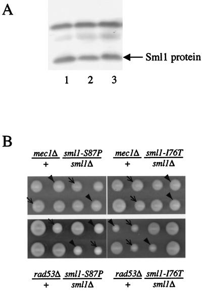
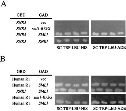
In budding yeast, MEC1 and RAD53 are essential for cell growth. Previously we reported that mec1 or rad53 lethality is suppressed by removal of Sml1, a protein that binds to the large subunit of ribonucleotide reductase (Rnr1) and inhibits RNR activity. To understand further the relationship between this suppression and the Sml1-Rnr1 interaction, we randomly mutagenized the SML1 open reading frame. Seven mutations were identified that did not affect protein expression levels but relieved mec1 and rad53 inviability. Interestingly, all seven mutations abolish the Sml1 interaction with Rnr1, suggesting that this interaction causes the lethality observed in mec1 and rad53 strains. The mutant residues all cluster within the 33 C-terminal amino acids of the 104-amino-acid-long Sml1 protein. Four of these residues reside within an alpha-helical structure that was revealed by nuclear magnetic resonance studies. Moreover, deletions encompassing the N-terminal half of Sml1 do not interfere with its RNR inhibitory activity. Finally, the seven sml1 mutations also disrupt the interaction with yeast Rnr3 and human R1, suggesting a conserved binding mechanism between Sml1 and the large subunit of RNR from different species.


Figures

FIG. 1
FIG. 1
Isolation of loss-of-function sml1 mutations. (A) Scheme for isolating loss-of-function sml1 mutations. The SML1 ORF was mutagenized by PCR amplification (see Materials and Methods). The PCR fragments were cotransformed into yeast strain U1047 (MATa ade2Δ ade3Δ sml1Δ::HIS3 mec1-1 [pC87 ADE3-URA3-MEC1]) with gapped vector pACTII that has a GAD fused with an HA tag (pGAD-HA). Strain U1047 forms solid red colonies when the fusion protein contains wild-type SML1 (pGAD-HA-SML1); it forms red-white sectored colonies when the fusion protein contains loss-of-function sml1 mutations (pGAD-HA-sml1) or only the GAD-HA fusion. (B) Expression of the mutated Sml1 proteins. The GAD-HA fusion proteins containing wild-type and mutated Sml1 were detected on a protein blot by using anti-HA antibody (12CA5). The bracket indicates the GAD-HA-Sml1 proteins. The multiple bands are due to phosphorylation that is stabilized in GAD-HA-Sml1 fusion proteins (data not shown). (C) Positions of the seven sml1 mutations. The amino acid (a.a.) changes and their positions on the protein are illustrated. The hatched boxes depict alpha-helical regions revealed by NMR.
FIG. 2
FIG. 2
sml1 mutations rescue mec1 and rad53 lethality. (A) The Sml1 protein level in wild-type (lane 1), sml1-I76T (lane 2), and sml1-S87P (lane 3) strains was examined by protein blotting. The arrow indicates the Sml1 protein. The darker band above Sml1 cross-reacts to anti-Sml1 serum and was used as a loading control. (B) Tetrad analysis of sml1-I76T and sml1-S87P. The genotype of the diploid is above or below each panel. Two tetrads are shown for each and are displayed horizontally. The arrows (➞) indicate mec1Δ sml1Δ (top) and rad53Δ sml1Δ (bottom). The arrowheads (▾) indicate mec1Δ sml1-S87P (top left), mec1Δ sml1-I76T (top right), rad53Δ sml1-S87P (bottom left), and rad53Δ sml1-I76T (bottom right). In all cases, sml1Δ and two sml1 mutations suppressed mec1Δ and rad53Δ to a similar degree.
FIG. 3
FIG. 3
Interaction between SML1 or sml1 mutants and RNR1 or DUN1 in two-hybrid assays. (A) Plasmids containing GAD-HA-SML1 or GAD-HA-sml1-R72G were cotransformed with plasmids containing GBD vector alone (vec), GBD-DUN1, or GBD-RNR1 into two-hybrid strain PJ69-4A. The interactions were assayed by activation of the HIS3 reporter on SC-TRP-LEU-HIS medium. Four or five transformants are shown for each cotransformation. sml1-R72G failed to bind to Rnr1 but still interacted with Dun1. Similar observations were made for the other six sml1 mutations (data not shown). (B) LacZ activity was measured in Miller Units (1). The white bars represent the interactions between sml1 mutants and Rnr1, and the black bars represent those between sml1 mutants and the empty GBD vector. WT, wild type.
FIG. 4
FIG. 4
The sml1 mutations abolish the interaction with RNR3 and human R1. Different sets of plasmids were cotransformed into two-hybrid strain PJ69-4A, and the activation of HIS3 and ADE2 reporters were examined by growing transformants on SC-TRP-LEU-HIS and SC-TRP-LEU-ADE plates, respectively. (A) The GBD-RNR3 plasmid was cotransformed into PJ69-4A with plasmids containing either GAD (vec), GAD-sml1-R72G, GAD-SML1, or GAD-RNR1. (B) The GBD-human R1 plasmid was cotransformed into two-hybrid strain PJ69-4A with plasmids containing either GAD (vec), GAD-SML1, or GAD-sml1-R72G. GBD-RNR1 and GAD-SML1 are also included for comparison.
FIG. 5
FIG. 5
Secondary Cα chemical shifts of individual amino acid residues in Sml1 determined from NMR data. Positive Cα values indicate the presence of an alpha helix (shown by dashed lines). The positions of the seven sml1 mutations are labeled.
FIG. 6
FIG. 6
Effect of Sml1 mutations on its inhibitory activity of RNR. Assay mixtures contained 20 mM HEPES-KOH (pH 7.4), 200 mM potassium acetate, 5 mM ATP, 20 mM magnesium acetate, 1 mM [3H]CDP (specific activity, 27,000 cpm/nmol), 20 μM FeCl3, 20 mM dithiothreitol, 0.2 μM Rnr1, 1 μM Rnr2/Rnr4 heterodimer, and the indicated concentrations of recombinant wild-type or mutated Sml1. The reaction mixtures were incubated at 30°C for 20 min in a final volume of 50 μl. At least two independent assays were performed for each concentration of wild-type and mutant Sml1. After incubation, the samples were processed as described earlier to obtain the amount of dCDP formed (4). Shown are wild-type Sml1 (WT) (open diamond), Δ28-50 (filled diamond), Δ2-39 (open square), S75A (open circle), S75P (open triangle), R72A (filled circle), F104L (filled square), and L73A (filled triangle).

Similar articles

Cited by

References

    1. Adams A, Gottschling D E, Kaiser C A, Stearns T. Methods in yeast genetics, a Cold Spring Harbor Laboratory course manual. Cold Spring Harbor, N.Y: Cold Spring Harbor Laboratory Press; 1997.
    1. Bell D W, Varley J M, Szydlo T E, Kang D H, Wahrer D C, Shannon K E, Lubratovich M, Verselis S J, Isselbacher K J, Fraumeni J F, Birch J M, Li F P, Garber J E, Haber D A. Heterozygous germ line hCHK2 mutations in Li-Fraumeni syndrome. Science. 1999;286:2528–2531. - PubMed
    1. Caspari T. How to activate p53. Curr Biol. 2000;10:R315–R317. - PubMed
    1. Chabes A, Domkin V, Larsson G, Liu A, Graslund A, Wijmenga S, Thelander L. Yeast ribonucleotide reductase has a heterodimeric iron-radical-containing subunit. Proc Natl Acad Sci USA. 2000;97:2474–2479. - PMC - PubMed
    1. Chabes A, Domkin V, Thelander L. Yeast Sml1, a protein inhibitor of ribonucleotide reductase. J Biol Chem. 1999;274:36679–36683. - PubMed

Publication types

MeSH terms

LinkOut - more resources