. 2000 Nov 14;39(45):13760-71.
doi: 10.1021/bi001472t.

Identification of the Cu2+ binding sites in the N-terminal domain of the prion protein by EPR and CD spectroscopy

Affiliations

Identification of the Cu2+ binding sites in the N-terminal domain of the prion protein by EPR and CD spectroscopy

E Aronoff-Spencer et al. Biochemistry. .

Abstract

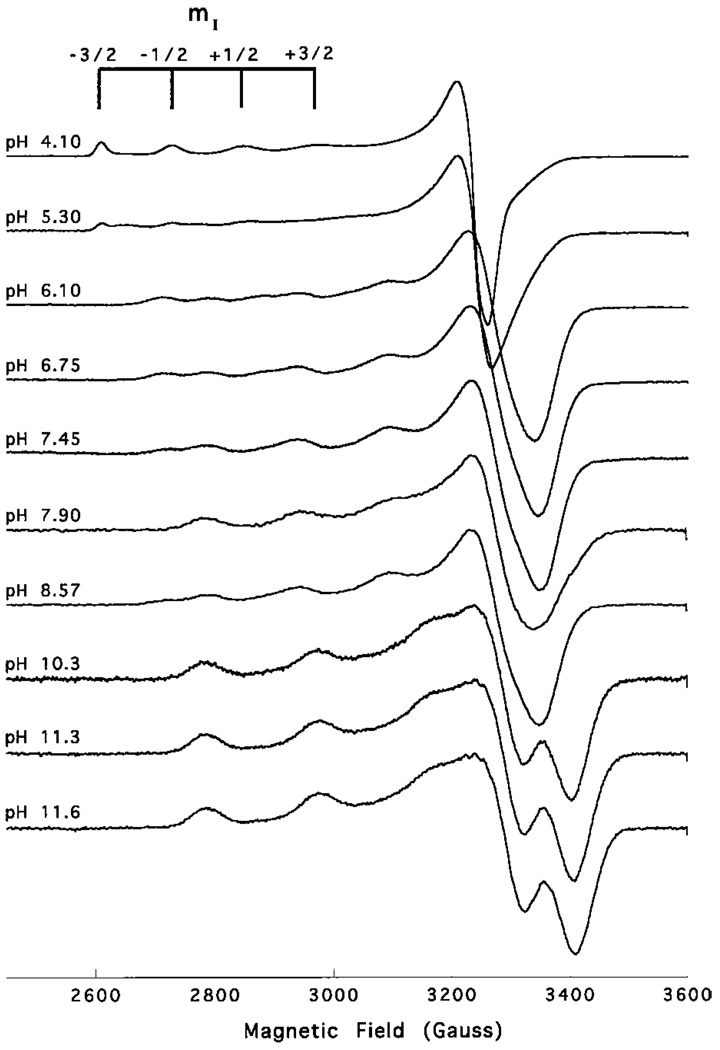
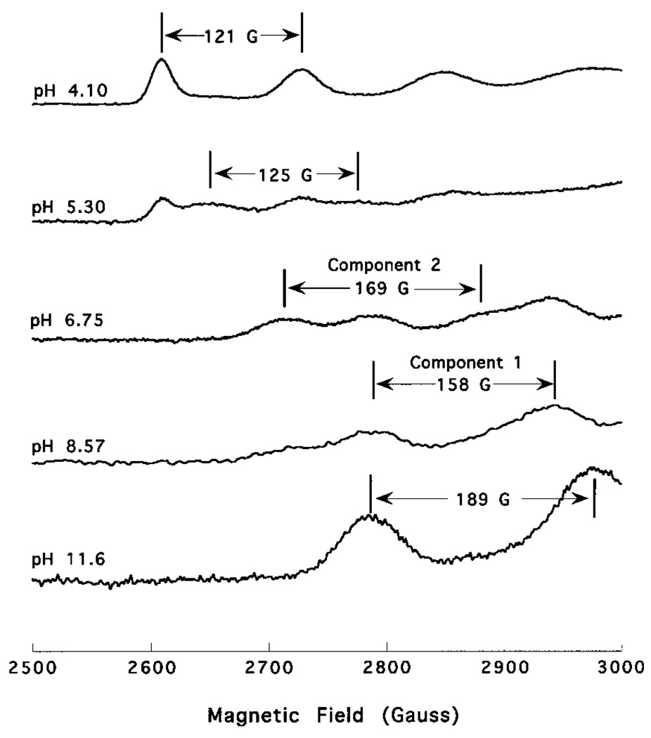
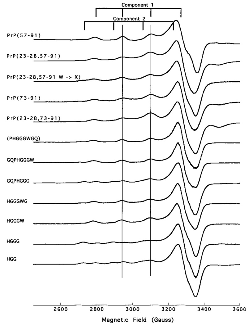
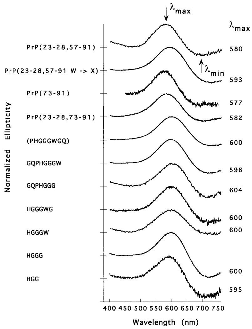
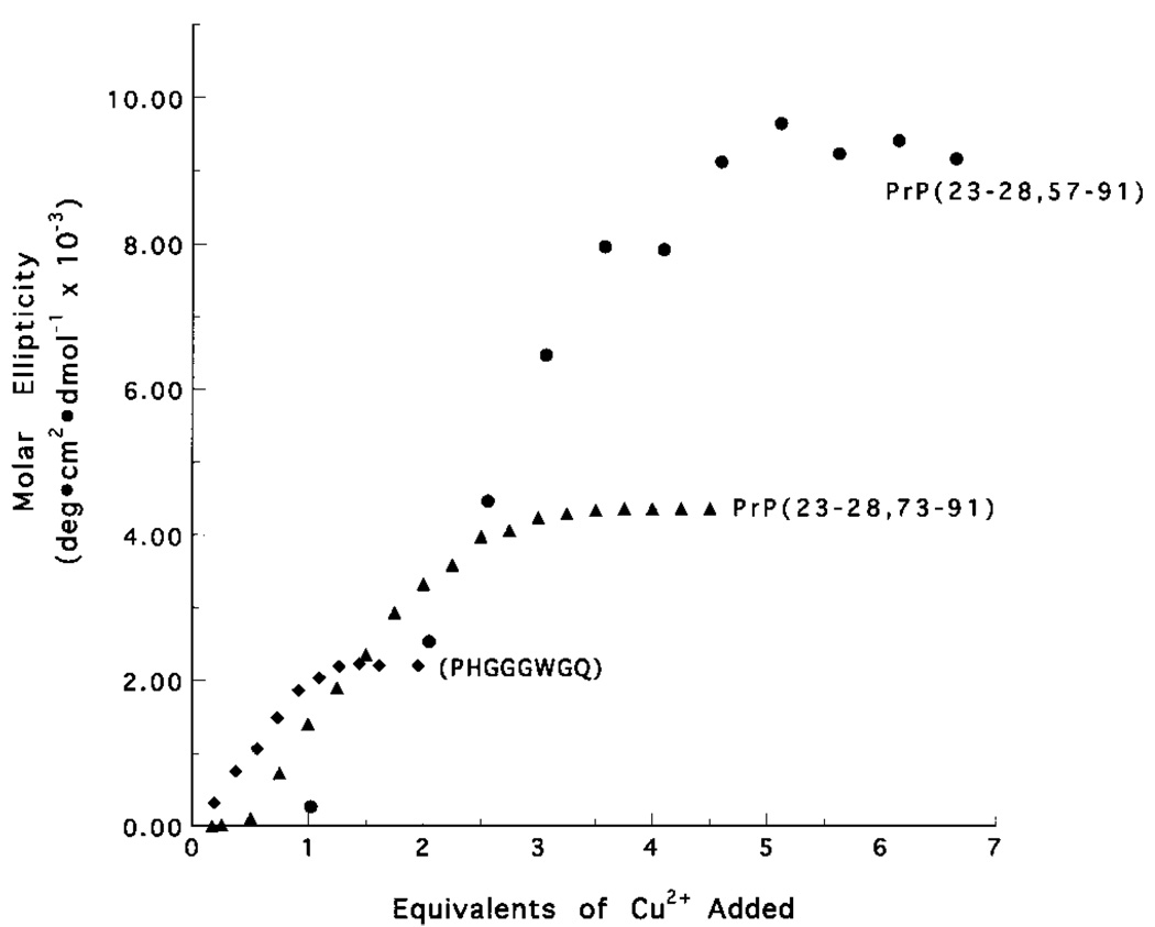
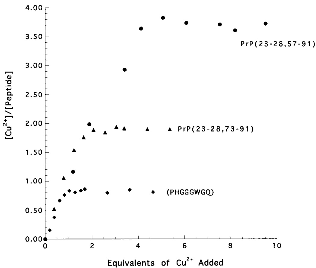
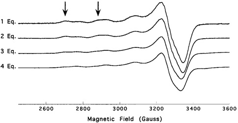
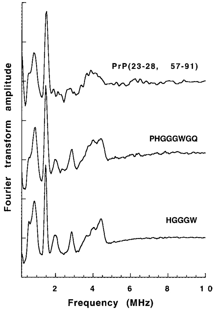
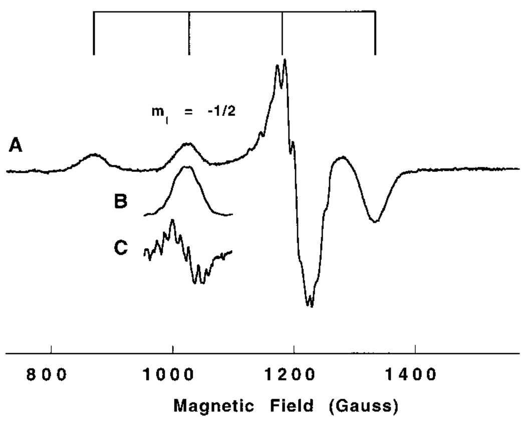
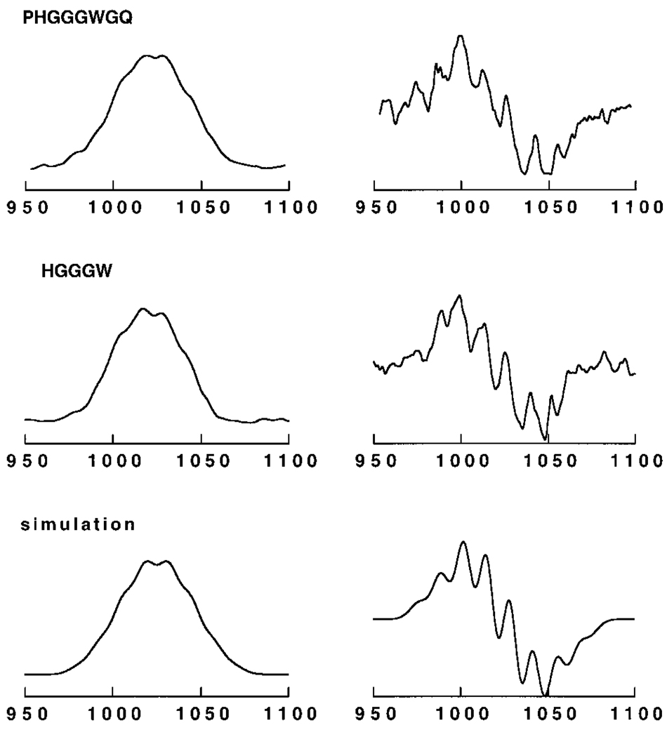
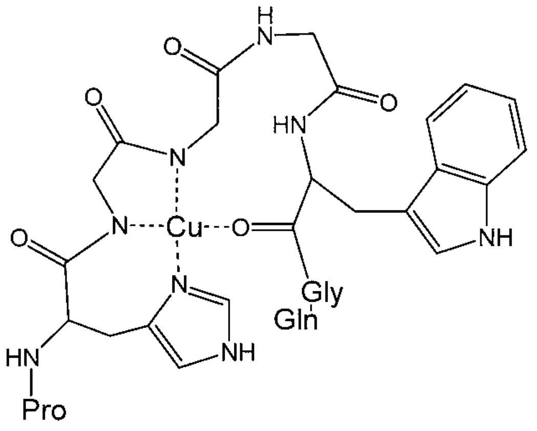
Recent evidence indicates that the prion protein (PrP) plays a role in copper metabolism in the central nervous system. The N-terminal region of human PrP contains four sequential copies of the highly conserved octarepeat sequence PHGGGWGQ spanning residues 60-91. This region selectively binds divalent copper ions (Cu(2+)) in vivo. To elucidate the specific mode and site of binding, we have studied a series of Cu(2+)-peptide complexes composed of 1-, 2-, and 4-octarepeats and several sub-octarepeat peptides, by electron paramagnetic resonance (EPR, conventional X-band and low-frequency S-band) and circular dichroism (CD) spectroscopy. At pH 7.45, two EPR active binding modes are observed where the dominant mode appears to involve coordination of three nitrogens and one oxygen to the copper ion, while in the minor mode two nitrogens and two oxygens coordinate. ESEEM spectra demonstrate that the histidine imidazole contributes one of these nitrogens. The truncated sequence HGGGW gives EPR and CD that are indistinguishable from the dominant binding mode observed for the multi-octarepeat sequences and may therefore comprise the fundamental Cu(2+) binding unit. Both EPR and CD titration experiments demonstrate rigorously a 1:1 Cu(2+)/octarepeat binding stoichiometry regardless of the number of octarepeats in a given peptide sequence. Detailed spin integration of the EPR signals demonstrates that all of the bound Cu(2+) is detected thereby ruling out strong exchange coupling that is often found when there is imidazolate bridging between paramagnetic metal centers. A model consistent with these data is proposed in which Cu(2+) is bound to the nitrogen of the histidine imidazole side chain and to two nitrogens from sequential glycine backbone amides.


Figures

Figure 1
Figure 1
X-band EPR spectra of 63Cu2+ loaded PrP(23–28, 57–91) at different pH values. The grid at top identifies the four hyperfine lines arising from coupling to the 63Cu (I = 3/2) nucleus. All spectra were collected at 77 K, νo = 9.43 GHz, and a sweep width of 1200 G.
Figure 2
Figure 2
Expansion of the low field (parallel) region from Figure 1 at selected pH values. As the pH increases there are marked changes of the four hyperfine lines reflecting changes in the ligand environment of the copper ion. Distinct species apparent at various points in the pH range are indicated.
Figure 3
Figure 3
X-band EPR spectra of all 63Cu2+/peptide complexes at pH values 7.45 ± 0.07. Spectral component 1 and component 2 are indicated with grids at top. Vertical lines have been added to the mI = −1/2 and mI = +1/2 hyperfine lines of component 1 to aid in comparison of g and A values between complexes. All spectra collected at 77 K, νo = 9.43 GHz, and a sweep width of 1200 G.
Figure 4
Figure 4
Visible region CD spectra of all Cu2+/peptide complexes at pH 7.4. The spectra are normalized along the y axis for comparison of λmax and λmin. The approximate baseline for each spectrum is indicated by hatch marks at the left. The λmax of the positive CD band is listed to the right of each spectrum. Because of poor solubility, PrP(57–91) is not included in this figure. All spectra collected at 298 K.
Figure 5
Figure 5
CD signal intensity at λmax as a function of titrated Cu2+ at pH 7.4 for the octarepeat, PrP(23–28, 73–91), and PrP(23–28, 57–91). Peptide concentrations are 398, 333, and 51 µM, respectively, and copper ion concentrations are reported in equivalents per peptide.
Figure 6
Figure 6
Integrated EPR signal intensity as a function of titrated Cu2+ at pH 7.4 for the octarepeat, PrP(23–28, 73–91), and PrP-(23–28, 57–91). Peptide concentrations are 1000, 500, and 250 µM, respectively, and the copper ion concentration is reported in equivalents per peptide. All spectra collected at 298 K.
Figure 7
Figure 7
X-band EPR spectra of 133 µM PrP(23–28, 57–91) with 1, 2, 3, and 4 equiv of Cu2+ at pH 7.4. Each spectrum has been normalized to unit area. Arrows have been added above the mI = −3/2 and mI = −1/2 hyperfine lines to highlight changes in this region. All spectra collected at 77 K, νo = 9.43 GHz, and a sweep width of 1200 G.
Figure 8
Figure 8
Three pulse ESEEM spectra for PrP(23–28, 57–91), the octarepeat (PHGGGWGQ) and HGGGW obtained at X-band. Spectra were obtained at 4.2 K from the g region of the spectrum with τ = 150 ns.
Figure 9
Figure 9
S-band EPR (3.5 GHz) of the octarepeat (PHGGG-WGQ). The grid at the top shows the 63Cu hyperfine splitting, and the mI = −1/2 line is indicated. Trace A is the full field scan; trace B is the mI = −1/2 line scanned with high resolution and signal averaging; trace C is the derivative of B emphasizing the multiplet structure.
Figure 10
Figure 10
Expansion of the mI = −1/2 lines of the S-band EPR spectra. The octarepeat (PHGGGWGQ) and HGGGW lines are shown (left) along with their derivatives (right). The simulated spectrum is from a model of three equivalent nitrogens (aN = 13 G) and one proton (aH = 10 G).
Figure 11
Figure 11
Proposed bond-line model of the dominant binding mode (component 1) of the octarepeat segment (PHGGGWGQ).

References

    1. Prusiner SB. Science. 1982;216:136–144. - PubMed
    1. Prusiner SB. Science. 1991;252:1515–1522. - PubMed
    1. Prusiner SB. Science. 1997;278:245–251. - PubMed
    1. Zahn R, Liu A, Luhrs T, Riek R, von Schroetter C, Lopez Garcia F, Billeter M, Calzolai L, Wider G, Wuthrich K. Proc. Natl. Acad. Sci. U.S.A. 2000;97:145–150. - PMC - PubMed
    1. Riek R, Hornemann S, Wider G, Billeter M, Glockshuber R, Wuthrich K. Nature. 1996;382:180–182. - PubMed

Publication types