. 2000 Nov 13;151(4):779-88.
doi: 10.1083/jcb.151.4.779.

Differential expression of lumican and fibromodulin regulate collagen fibrillogenesis in developing mouse tendons

Affiliations

Differential expression of lumican and fibromodulin regulate collagen fibrillogenesis in developing mouse tendons

Y Ezura et al. J Cell Biol. .

Abstract

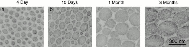
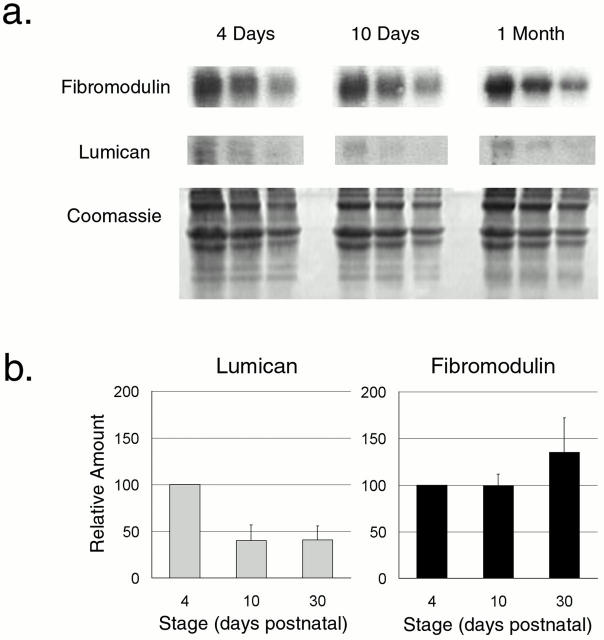
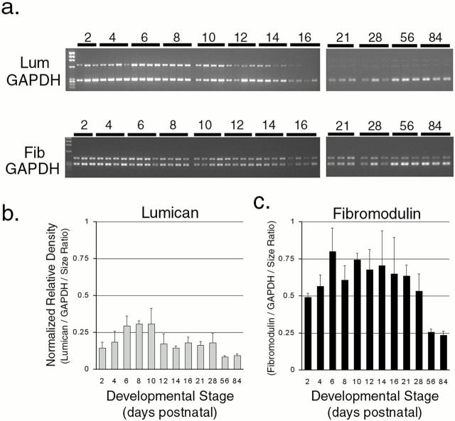
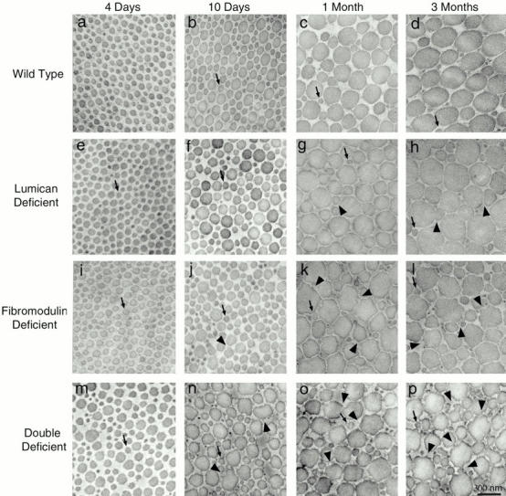
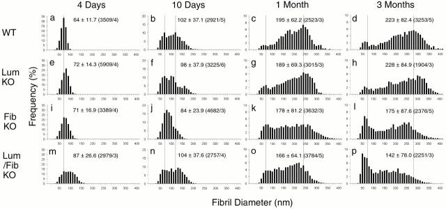
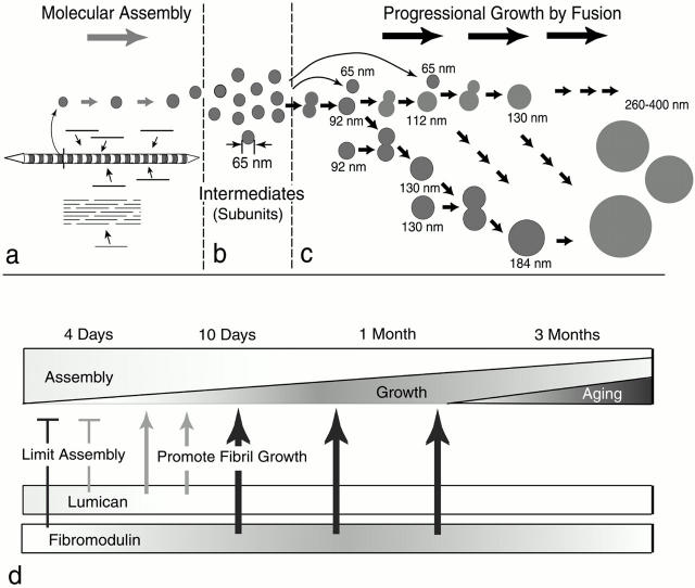
Collagen fibrillogenesis is finely regulated during development of tissue-specific extracellular matrices. The role(s) of a leucine-rich repeat protein subfamily in the regulation of fibrillogenesis during tendon development were defined. Lumican-, fibromodulin-, and double-deficient mice demonstrated disruptions in fibrillogenesis. With development, the amount of lumican decreases to barely detectable levels while fibromodulin increases significantly, and these changing patterns may regulate this process. Electron microscopic analysis demonstrated structural abnormalities in the fibrils and alterations in the progression through different assembly steps. In lumican-deficient tendons, alterations were observed early and the mature tendon was nearly normal. Fibromodulin-deficient tendons were comparable with the lumican-null in early developmental periods and acquired a severe phenotype by maturation. The double-deficient mice had a phenotype that was additive early and comparable with the fibromodulin-deficient mice at maturation. Therefore, lumican and fibromodulin both influence initial assembly of intermediates and the entry into fibril growth, while fibromodulin facilitates the progression through growth steps leading to mature fibrils. The observed increased ratio of fibromodulin to lumican and a competition for the same binding site could mediate these transitions. These studies indicate that lumican and fibromodulin have different developmental stage and leucine-rich repeat protein specific functions in the regulation of fibrillogenesis.


Figures

Figure 1
Figure 1
Collagen fibril structure during development in wild-type mouse tendons. Transmission electron micrographs of transverse sections from mouse flexor tendons from normal mice (a–d). Fibril structure was analyzed at different developmental stages, 4 d (a), 10 d (b), 1 mo (c), and 3 mo (d) postnatal. Bar, 300 nm.
Figure 3
Figure 3
Lumican and fibromodulin content during normal tendon development. (a) A representative semiquantitative Western analysis of lumican and fibromodulin during development in the normal mouse tendon is presented. Tendons were extracted in 4 M guanidine-HCl at 4 d, 10 d, and 1 mo. 80, 40, and 20 μg of total protein from each time point were loaded onto the gel. The core proteins were transferred, reacted with antilumican or antifibromodulin antisera followed by radiolabeled goat anti–rabbit IgG, and quantitated using phosphoimaging. A duplicate gel stained with Coomassie shows similar amounts of type I collagen in the extracts. (b) The relative lumican and fibromodulin content in the tendon at 4 d, 10 d, and 1 mo postnatal were derived from three independent experiments. The mean values for both lumican and fibromodulin were set to 100 at 4 d and the results were plotted as a function of development (bars indicate SD).
Figure 2
Figure 2
Expression of lumican and fibromodulin during normal tendon development. Lumican and fibromodulin expression were analyzed using semiquantitative RT-PCR. (a) Representative ethidium bromide stained gels showing PCR product bands for lumican, fibromodulin, and coamplified GAPDH bands. Three or four different batches of PCR products generated from independently prepared total RNA between postnatal day 4 and 16 or day 21 and 3 mo were applied to a single gel. (b and c) Band density was determined densitometrically and relative density was obtained by using both band density and the PCR product size ratio, lumican 656 bp, fibromodulin 418 bp, and GAPDH 289 bp. The relative density was normalized using GAPDH and plotted for (b) lumican and (c) fibromodulin.
Figure 4
Figure 4
Localization of lumican and fibromodulin core proteins in normal tendon development. Immunofluorescence microscopy showing the localization of lumican (a) and fibromodulin (b) core proteins in longitudinal sections of normal mouse tendons at 10 d postnatal. Control sections (c) were treated identically without the primary antibody. Bar, 50 μm.
Figure 5
Figure 5
Collagen fibril structure during development in normal wild-type, lumican-, fibromodulin-, and double lumican/fibromodulin–deficient mice. Transmission electron micrographs of transverse sections from mouse flexor tendons from normal mice (a–d) and mutant mice (e–p). Fibril structure was analyzed at different developmental stages between 4 d and 3 mo postnatal, 4 d (a, e, i, and m), 10 d (b, f, j, and n), 1 mo (c, g, k, and o), and 3 mo (d, h, l, and p) postnatal. Arrows indicate fibrils with diameters of ∼64 nm, the diameter seen in normal 4-d postnatal tendons. Arrowheads indicate irregular fibril profiles. Bar, 300 nm.
Figure 6
Figure 6
Collagen fibril diameter distributions during development of wild-type, lumican-null, fibromodulin-null, and double lumican/fibromodulin–null tendons. Collagen fibril diameter distributions are presented as histograms for wild-type (a–d), lumican-deficient (e–h), fibromodulin-deficient (i–l), and double lumican/fibromodulin–deficient (m–p) mouse tendons. Vertical dotted lines indicate peaks observed in wild-type mouse tendons at ∼65, 120, 240, and 280 nm. (a–d) Means, SD, and number of fibrils measured (n)/number of different animals (n a) are presented as mean ± SD (n/n a) in the graphs.
Figure 7
Figure 7
Model for regulation of fibril growth by lumican and fibromodulin. The steps in collagen fibrillogenesis during tendon development are presented (a–c). (a) In the early steps of fibril formation, the molecular assembly of collagen monomers into fibril intermediates occurs in the pericellular space. Collagen molecules (bars) assemble into quarter-staggered arrays forming fibril intermediates, seen here as striated structures with tapered ends in longitudinal section and in cross section as circular profiles. Growth in length and diameter is by accretion of collagen at this stage. (b) Fibril intermediates ∼65 nm in mouse tendons are stabilized, presumably through their interactions with fibril-associated macromolecules. (c) The fibril intermediates are the basic units used in the growth of fibrils. Fusion of the fibril intermediates generates the mature fibril in a multistep manner. Progression through this growth process could be both by additive fusion (i.e., the 64-nm diameter intermediate adds to its product, indicated by horizontal arrows) and by like-fusion (i.e., products from different steps can only fuse with like products, indicated by arrows in the oblique direction). (d, top) The proportion of the assembly and growth steps occurring during development is illustrated. At early stages, assembly is the main event and its proportion gradually decreases to the minimum degree necessary for maintenance at maturation. The proportion of progressional growth increases gradually to ∼1 mo and decreases to maturation. (bottom) The expression data for lumican and fibromodluin is illustrated. The phenotypes observed in mutant mice indicate stage-specific regulatory mechanisms. At 4 d, both lumican and fibromodulin limit the assembly of the collagen monomers (bars). Characteristic of 10 d, progressional growth begins, and changes in both lumican and fibromodulin promote the transition from assembly to fibril growth by fusion (thin arrows). At later stages, only fibromodulin promotes the progressional growth steps (thick arrows).

References

    1. Birk D.E., Hahn R.A., Linsenmayer C.Y., Zycband E.I. Characterization of fibril segments from chicken embryo cornea, dermis and tendon. Matrix Biol. 1996;15:111–118. - PubMed
    1. Birk D.E., Linsenmayer T.F. Collagen fibril assembly, depostion, and organization into tissue-specific matrices. In: Yurchenco P.D., Birk D.E., Mecham R.P., editors. Extracellular Matrix Assembly and Structure. Academic Press, Inc; New York, NY: 1994. pp. 91–128.
    1. Birk D.E., Mayne R. Localization of collagen types I, III and V during tendon development. Changes in collagen types I and III are correlated with changes in fibril diameter. Eur. J. Cell Biol. 1997;72:352–361. - PubMed
    1. Birk D.E., Nurminskaya M.V., Zycband E.I. Collagen fibrillogenesis in situfibril segments undergo post-depositional modifications resulting in linear and lateral growth during matrix development. Dev. Dyn. 1995;202:229–243. - PubMed
    1. Birk D.E., Trelstad R.L. Extracellular compartments in tendon morphogenesiscollagen fibril, bundle, and macroaggregate formation. J. Cell Biol. 1986;103:231–240. - PMC - PubMed

Publication types

MeSH terms