. 2000 Nov 13;151(4):801-10.
doi: 10.1083/jcb.151.4.801.

c-Jun activation-dependent tumorigenic transformation induced paradoxically by overexpression or block of S-adenosylmethionine decarboxylase

Affiliations

c-Jun activation-dependent tumorigenic transformation induced paradoxically by overexpression or block of S-adenosylmethionine decarboxylase

A Paasinen-Sohns et al. J Cell Biol. .

Abstract

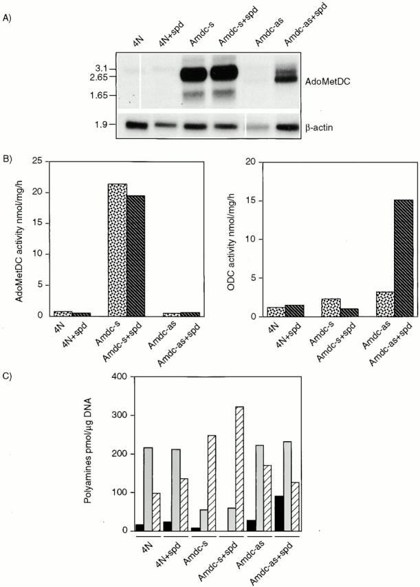
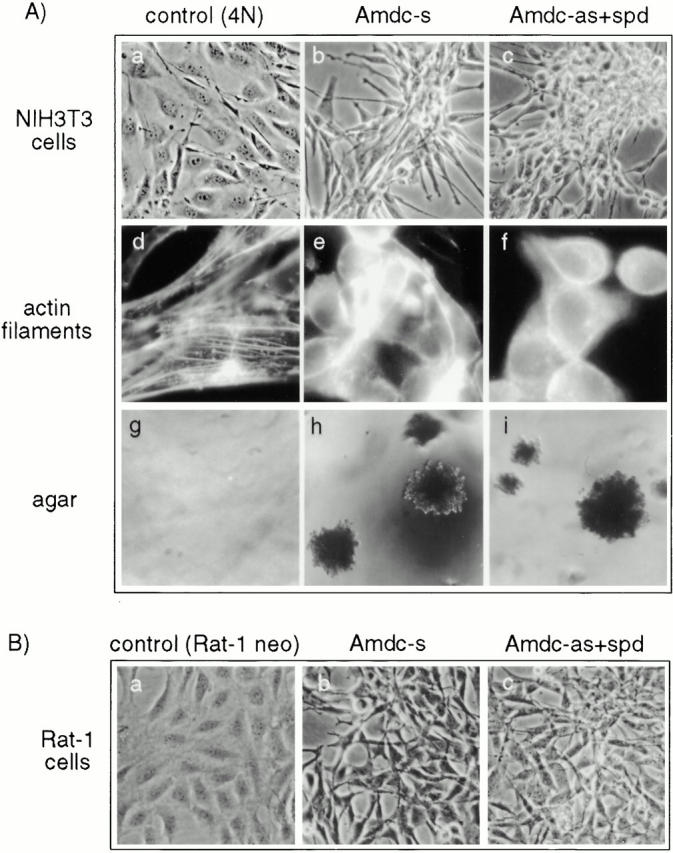
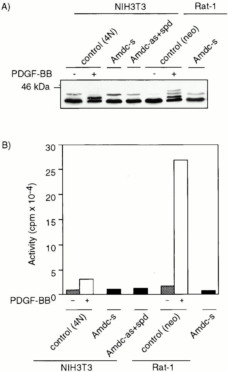
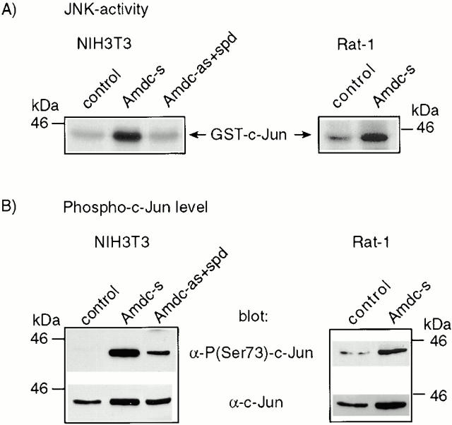
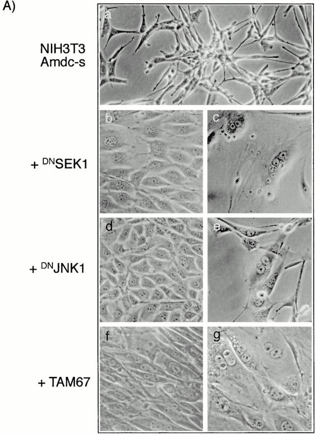
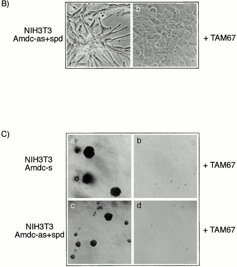
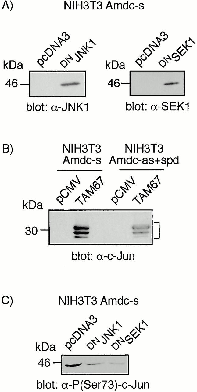
All mammalian cells absolutely require polyamines (putrescine, spermidine, and spermine) for growth. Here we show that the overexpression of cDNA for S-adenosylmethionine decarboxylase (AdoMetDC), the main regulatory enzyme in the biosynthesis of higher polyamines, induces transformation of rodent fibroblasts when expressed in the sense or the antisense orientation. Both transformants were able to induce invasive tumors in nude mice. Neither transformation was associated with activation of the mitogen-activated protein kinases Erk1 and Erk2. Instead, the AdoMet DC sense, but not antisense, transformants displayed constitutive activation of the c-Jun NH(2)-terminal kinase (JNK) pathway. However, both transformations converged on persistent phosphorylation of endogenous c-Jun at Ser73. The phenotype of the AdoMetDC sense transformants was reversed by expression of dominant-negative mutants of SEK1 (MKK4), JNK1, and c-Jun (TAM-67), which were also found to impair cytokinesis. Similarly, TAM-67 reverted the morphology of the AdoMetDC-antisense expressors. This report is the first demonstration of a protein whose overexpression or block of synthesis can induce cell transformation. In addition, we show that the polyamine biosynthetic enzymes require c-Jun activation for eliciting their biological effects.


Figures

Figure 2
Figure 2
Expression of human AdoMetDC cDNA, the activities of AdoMetDC and ODC, and polyamine contents in NIH3T3 transfectants are shown. (A) The Northern blot shows the expression levels of AdoMetDC mRNA in transfected cells. The integrity and loading of RNA was controlled by hybridizing the blot with human β-actin c-DNA. Note, the consistent lower β-actin signal in the Amdc-as cells may also reflect their retarded growth rate. The size of the mRNAs are indicated in kb on the left. (B) AdoMetDC (left) and ODC (right) activities were measured as described in Materials and Methods. Dotted bars, cells grown without spermidine addition; striped bars, cells grown with spermidine. (C) The amount of polyamines is expressed as pmol/μg DNA. Acetylated polyamines were not detected. Black bars, putrescine; gray bars, spermidine; striped bars, spermine. The enzymatic activities and the polyamine contents were determined from parallel dishes after 2 d of culture. The results are representative of four independent experiments.
Figure 1
Figure 1
(A) Morphology, actin filaments, and soft agar growth of NIH3T3 cells overexpressing human AdoMetDC cDNA in sense (Amdc-s) and antisense (Amdc-as) orientations are shown. (a, d, and g) Parental NIH3T3 cells transfected with the neomycin resistance gene and the empty pLTRpoly vector (control, 4N), (b, e, and h) Amdc-s cells and (c, f, and i) Amdc-as cells grown with 1 μM spermidine (Amdc-as+spd) in tissue cultures (a–c), stained for actin filaments (d–f), and grown in soft agar (g–i). (B) Morphology of Rat-1 cell transfectants. (a–c) Rat-1 control cells and transfectants expressing AdoMetDC sense and antisense constructs. Rat-1 Amdc-as cells were grown in the presence of spermidine, as described above.
Figure 3
Figure 3
Erk1 and Erk2 are not constitutively activated in AdoMetDC transformants. (A) Western blot analysis of the phosphorylation status of Erks. The electrophoretic mobilities of Erk1 and Erk2 in Amdc-s or Amdc-as+spd cells did not display upshifts of protein bands (in 12.5% SDS-PAGE) that were seen in control cells (4N and Rat-1) stimulated with PDGF-BB. (B) In vitro immunocomplex kinase assays showed a clear stimulation of MAPK activity in PDGF-BB–stimulated control cells, but no constitutive upregulation was detected in AdoMetDC transformants. Gray bars, cells starved for 24 h in 0.5% serum; white bars, starved cells stimulated with PDGF-BB for 15 min at 37°C; black bars, cells grown normally in 5% serum.
Figure 4
Figure 4
Endogenous JNK is constitutively activated in AdoMetDC-overexpressing NIH3T3 and Rat-1 cells, resulting in increased phosphorylation of c-Jun at Ser73. (A) The JNKs were purified by virtue of their binding to agarose-conjugated GST-c-Jun. The autoradiograms show the phosphorylation status of GST-c-Jun used as the substrate in the solid-phase kinase assays. (B) Western blots from the nuclear extracts show a strong phosphorylation of c-Jun at Ser73 in AdoMetDC transformants. The bottom rows show the total amount of c-Jun in nuclear extracts. The levels of JunD and ATF-2 remained constant and were used as loading controls (data not shown).
Figure 5
Figure 5
The transformed morphology of AdoMetDC-overexpressing cells is reversed by the expression of dominant-negative mutants of SEK1, JNK1, and c-Jun (TAM-67). (A) The morphology of (a) Amdc-s cells was changed to a more flattened and normal-looking phenotype by expressing (b) DN SEK1, (d) DN JNK1, and (f) TAM67 and resulted in the formation of multinucleated, growth-arrested cell populations, indicative of cytokinetic failure, in all three transfections (c, e, and g). The cells were transfected with mutant plasmids and selection markers, grown in the presence of zeosin or puromycin for 10 d (b, d, and f) or 17–21 d (c, e, and g), and photomicrographed. (B) The transformed morphology of Amdc-as+spd cells is reversed by c-Jun mutant TAM67. The cells were transfected as described above. (C) The ability of the Amdc-s and Amdc-as+spd cells to grow in soft agar is inhibited by TAM67 expression. In both cell lines the inhibition of soft agar growth by TAM67 was >90%, and the colonies formed were tiny in size.
Figure 5
Figure 5
The transformed morphology of AdoMetDC-overexpressing cells is reversed by the expression of dominant-negative mutants of SEK1, JNK1, and c-Jun (TAM-67). (A) The morphology of (a) Amdc-s cells was changed to a more flattened and normal-looking phenotype by expressing (b) DN SEK1, (d) DN JNK1, and (f) TAM67 and resulted in the formation of multinucleated, growth-arrested cell populations, indicative of cytokinetic failure, in all three transfections (c, e, and g). The cells were transfected with mutant plasmids and selection markers, grown in the presence of zeosin or puromycin for 10 d (b, d, and f) or 17–21 d (c, e, and g), and photomicrographed. (B) The transformed morphology of Amdc-as+spd cells is reversed by c-Jun mutant TAM67. The cells were transfected as described above. (C) The ability of the Amdc-s and Amdc-as+spd cells to grow in soft agar is inhibited by TAM67 expression. In both cell lines the inhibition of soft agar growth by TAM67 was >90%, and the colonies formed were tiny in size.
Figure 6
Figure 6
The expression of DN JNK1, DN SEK1, and c-Jun (TAM67) mutant proteins and inhibition of c-Jun phosphorylation by DN JNK1 and DN SEK1. (A and B) Western blot analyses of the cell lysates for the expression of the mutant proteins. Note that there are multiple c-Jun TAM67 bands (marked with a bracket), as reported earlier (Brown et al. 1994). (C) Western blot analysis showing the inhibition of c-Jun phosphorylation in Amdc-s cells transfected with DN JNK1 and DN SEK1.

References

    1. Alani R., Brown P., Binétruy B., Dosaka H., Rosenberg R.K., Angel P., Karin M., Birrer M.J. The transactivating domain of the c-Jun proto-oncoprotein is required for cotransformation of rat embryo cells. Mol. Cell. Biol. 1991;11:6286–6295. - PMC - PubMed
    1. Alessandrini A., Greulich H., Huang W., Erikson R.L. Mek1 phosphorylation site mutants activate Raf-1 in NIH3T3 cells. J. Biol. Chem. 1996;271:31612–31618. - PubMed
    1. Angel P., Imagawa M., Chiu R., Stein B., Imbra R.J., Rahmsdorf H.J., Jonat C., Herrlich P., Karin M. Phorbol ester-inducible genes contain a common cis element recognized by a TPA-modulated trans-acting factor. Cell. 1987;49:729–739. - PubMed
    1. Attar B.M., Atten M.J., Holian O. MAPK activity is down-regulated in human colon adenocarcinomacorrelation with PKC activity. Anticancer Res. 1996;16:395–400. - PubMed
    1. Auvinen M., Paasinen A., Andersson L.C., Hölttä E. Ornithine decarboxylase activity is critical for cell transformation. Nature. 1992;360:355–358. - PubMed

Publication types

MeSH terms

Substances