. 2000 Nov 13;151(4):847-62.
doi: 10.1083/jcb.151.4.847.

Identification of ubiquilin, a novel presenilin interactor that increases presenilin protein accumulation

Affiliations

Identification of ubiquilin, a novel presenilin interactor that increases presenilin protein accumulation

A L Mah et al. J Cell Biol. .

Abstract

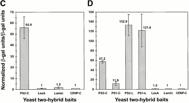
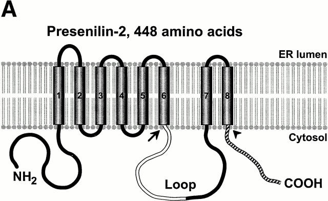
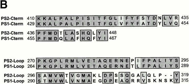
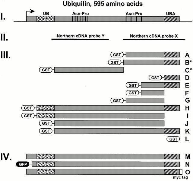
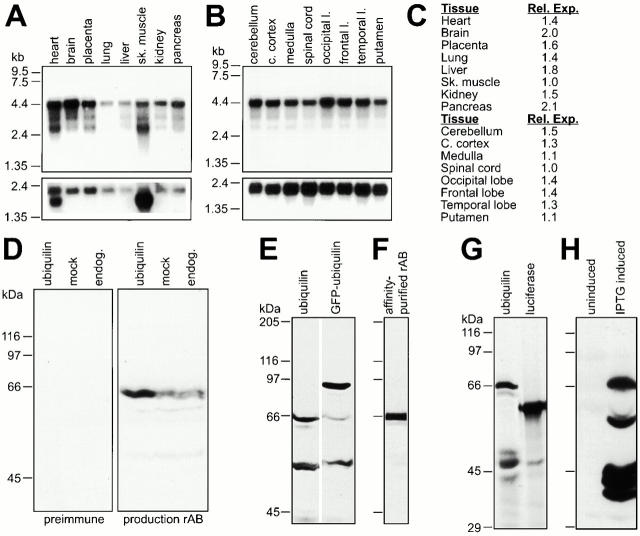
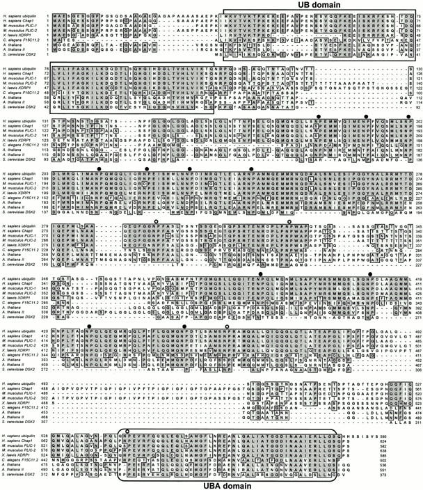
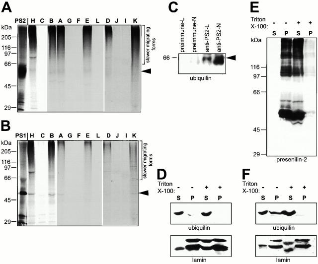
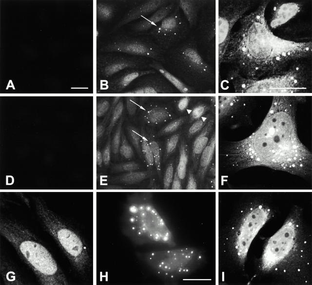
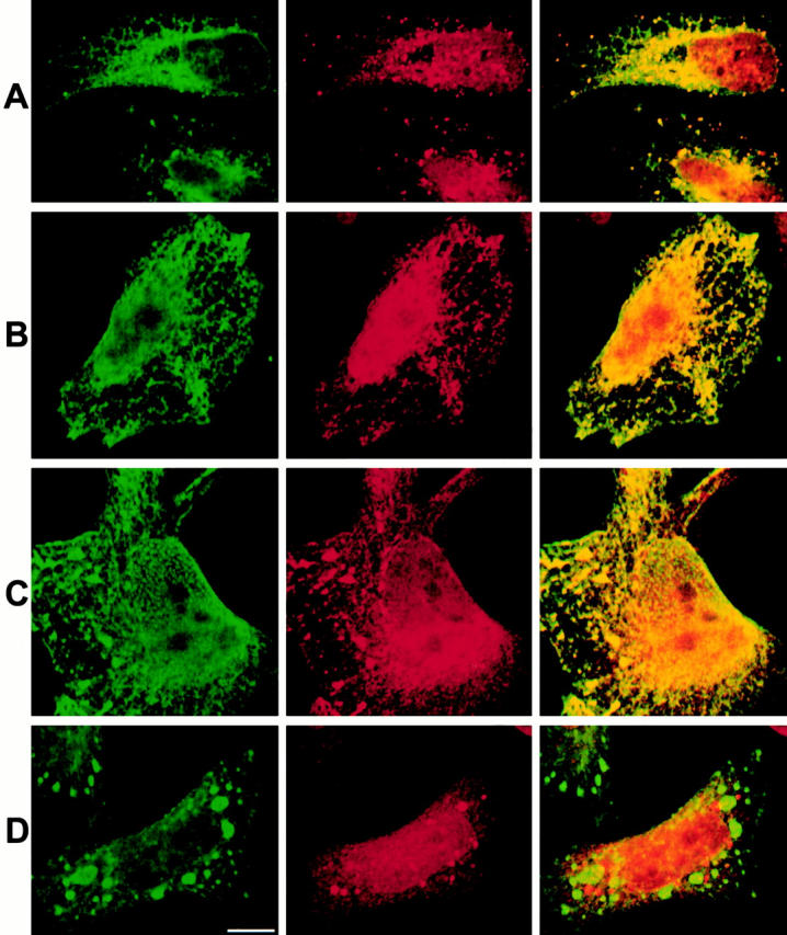
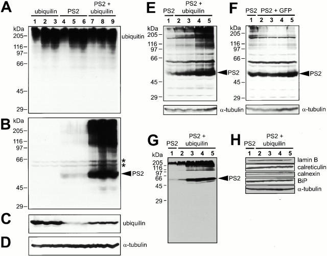
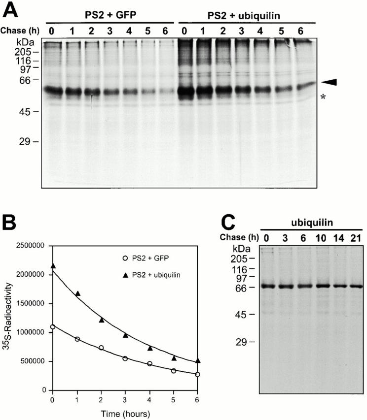
Mutations in the highly homologous presenilin genes encoding presenilin-1 and presenilin-2 (PS1 and PS2) are linked to early-onset Alzheimer's disease (AD). However, apart from a role in early development, neither the normal function of the presenilins nor the mechanisms by which mutant proteins cause AD are well understood. We describe here the properties of a novel human interactor of the presenilins named ubiquilin. Yeast two-hybrid (Y2H) interaction, glutathione S-transferase pull-down experiments, and colocalization of the proteins expressed in vivo, together with coimmunoprecipitation and cell fractionation studies, provide compelling evidence that ubiquilin interacts with both PS1 and PS2. Ubiquilin is noteworthy since it contains multiple ubiquitin-related domains typically thought to be involved in targeting proteins for degradation. However, we show that ubiquilin promotes presenilin protein accumulation. Pulse-labeling experiments indicate that ubiquilin facilitates increased presenilin synthesis without substantially changing presenilin protein half-life. Immunohistochemistry of human brain tissue with ubiquilin-specific antibodies revealed prominent staining of neurons. Moreover, the anti-ubiquilin antibodies robustly stained neurofibrillary tangles and Lewy bodies in AD and Parkinson's disease affected brains, respectively. Our results indicate that ubiquilin may be an important modulator of presenilin protein accumulation and that ubiquilin protein is associated with neuropathological neurofibrillary tangles and Lewy body inclusions in diseased brain.


Figures

Figure 1
Figure 1
Ubiquilin interacts with two different regions of presenilin proteins. (A) A schematic diagram of ER-bound human PS2 and shows eight transmembrane domains with its NH2 terminus, large hydrophilic loop, and COOH terminus all protruding into the cytoplasm. PS1 is believed to have a similar structure. The presenilin loop region (Loop, in white) and COOH terminus (striped) were used in Y2H assays. Two PS2 mutants used in this study contained progressively longer COOH-terminal deletions: PS2(ΔC), which terminated at the arrowhead, and PS2(ΔLC), which terminated at the arrow. (B) The amino acid sequence of PS1 and PS2 COOH terminus and loop regions that were used as Y2H baits. (C) Y2H β-galactosidase liquid culture interaction assay of an ubiquilin-prey clone (Fig. 2 III, construct B without GST) with PS2-COOH terminus (PS2-C), LexA alone, nuclear lamin B, and CENP-C baits. The β-galactosidase units were normalized to CENP-C, the bait with the weakest interaction. (D) β-Galactosidase interaction assay data of a near full-length ubiquilin-prey clone (Fig. 2 III, construct H without GST) repeated with the same baits described in C along with PS1-COOH (PS1-C) terminus, PS2-Loop (PS2-L), and PS1-Loop (PS1-L) baits. Again, β-galactosidase units were normalized to CENP-C.
Figure 1
Figure 1
Ubiquilin interacts with two different regions of presenilin proteins. (A) A schematic diagram of ER-bound human PS2 and shows eight transmembrane domains with its NH2 terminus, large hydrophilic loop, and COOH terminus all protruding into the cytoplasm. PS1 is believed to have a similar structure. The presenilin loop region (Loop, in white) and COOH terminus (striped) were used in Y2H assays. Two PS2 mutants used in this study contained progressively longer COOH-terminal deletions: PS2(ΔC), which terminated at the arrowhead, and PS2(ΔLC), which terminated at the arrow. (B) The amino acid sequence of PS1 and PS2 COOH terminus and loop regions that were used as Y2H baits. (C) Y2H β-galactosidase liquid culture interaction assay of an ubiquilin-prey clone (Fig. 2 III, construct B without GST) with PS2-COOH terminus (PS2-C), LexA alone, nuclear lamin B, and CENP-C baits. The β-galactosidase units were normalized to CENP-C, the bait with the weakest interaction. (D) β-Galactosidase interaction assay data of a near full-length ubiquilin-prey clone (Fig. 2 III, construct H without GST) repeated with the same baits described in C along with PS1-COOH (PS1-C) terminus, PS2-Loop (PS2-L), and PS1-Loop (PS1-L) baits. Again, β-galactosidase units were normalized to CENP-C.
Figure 2
Figure 2
Schematic drawings of ubiquilin expression constructs. (I) The full-length ubiquilin polypeptide consists of 595 residues and contains an NH2-terminal UB domain (speckled), a COOH-terminal UBA domain (striped), and several regularly spaced asparagine-proline (Asn-Pro) repeats (vertical bars). (II) The probes used in human Northern blots. (III) GST-fusion constructs: A (N393–S595 aa), B (Q378–S595 aa), C (Q113–M377 aa), D (Q541–S595 aa), E (D449–S595 aa), F (D449–L540 aa), G (N393–L540 aa), H (M37–S595 aa), I (M37–L540 aa), J (Q113–L540 aa), K (Q113–S595 aa), and L (GST alone). The ubiquilin portions of constructs A and B were isolated in the original Y2H screen. Bacterially expressed GST–fusion B and C polypeptides were used as immunogens for anti-ubiquilin pAb production (*). (IV) Mammalian expression constructs: M, full-length untagged ubiquilin; N, NH2-terminal GFP-tagged ubiquilin fused at residue 20 (Ala); and O, COOH-terminal myc epitope-tagged ubiquilin.
Figure 3
Figure 3
Ubiquilin mRNA and protein expression. (A) Human multiple tissue were analyzed by Northern blot and probed with ubiquilin cDNA fragment X (Fig. 2 II). After stripping, the blot was reprobed with a β-actin control fragment (shown below). (B) Northern blot of specific regions of the human brain probed with ubiquilin cDNA fragment Y (Fig. 2 II), with a reprobe with β-actin, which is shown below. (C) Quantification of ubiquilin mRNA expression levels. Relative expression of ubiquilin in different tissues was determined by densitometric analysis of the autoradiographs and relating the ubiquilin band intensities to the levels of β-actin hybridization from the same lanes. The values are presented after normalization against skeletal muscle (above) and spinal cord (below). (D–F) Characterization of anti-ubiquilin antibodies. Rabbit antibodies were raised against GST–ubiquilin fusion proteins B and C, generating anti-ubiquilin-B and anti–ubiquilin-C antibodies, respectively. (D) Anti–ubiquilin-B antibody was used to detect overexpressed and endogenous ubiquilin in ubiquilin transfected, mock-transfected, and untransfected (endogenous) HeLa cells. The anti–ubiquilin-B antibody detected a 66-kD doublet band, whereas preimmune sera did not. (E) Anti–ubiquilin-C antibody also recognized the 66-kD band in untransfected HeLa lysates and to varying extents a ∼55-kD band. HeLa cells transfected with GFP–ubiquilin (Fig. 2 IV, N) contain an additional 93-kD reactive band, due to the fusion of the 27-kD moiety of GFP with ubiquilin. (F) Affinity-purified anti–ubiquilin-C antibody specifically reacts with the 66-kD band from transfected HeLa cell lysates. (G) Full-length in vitro transcribed and translated [35S]methionine-radiolabeled human ubiquilin polypeptides migrated at 66 kD, whereas radiolabeled luciferase migrated at 61 kD. (H) Uninduced and IPTG-induced lysates of bacteria transformed with untagged full-length human ubiquilin and probed with anti–ubiquilin-B antibodies. Full-length immunoreactive ubiquilin (66 kD) and a series of smaller breakdown products are only seen in the induced lysate.
Figure 4
Figure 4
Ubiquilin shares significant homology with several other proteins. The inferred amino acid sequence of the human ubiquilin ORF and its homology to several related proteins: Homo sapiens Chap1, a protein involved in binding Hsp70-like Stch protein; Mus musculus PLIC-1 and PLIC-2, proteins involved in linking integrins to vimentin; X. laevis XDRP1, a protein which binds cyclin A; C. elegans F15C11.2 and two A. thaliana proteins, proteins with unknown functions; and S. cerevisiae DSK2, involved in spindle pole body duplication. Identical residues are darkly shaded, whereas similar residues are lightly shaded. Only homology in five or more sequences are shown. All the proteins contain UB domains (square box) and UBA domains (rounded box), which are extremely well conserved in sequence. In addition, the central region between the UB and UBA domains contain several asparagine-proline repeats, which are either regularly spaced apart (closed circles) or located elsewhere throughout the protein (open circles). The high overall homology of the proteins suggests that they belong to the same multigene family.
Figure 5
Figure 5
Ubiquilin binds presenilins in vitro. (A and B) GST pull-down experiments. Full-length in vitro synthesized 35S-labeled PS2 and PS1 (first lanes) migrated in SDS-PAGE gels with broad bands of 54 and 48 kD (arrowheads), respectively, along with a smear of slower migrating forms, presumably due to the highly hydrophobic nature of the proteins. [35S]PS complexes (especially the slower migrating forms) were retained by GST–ubiquilin constructs containing the UBA domain (lane letters correspond to constructs shown in Fig. 2 III), but not by those lacking the domain, or by GST alone. (C) Immunoprecipitation experiments. PS2-transfected HeLa cell lysates were immunoprecipitated with preimmune sera or corresponding anti–PS2-Loop antibody and anti-PS2 NH2 terminus antibody. After separation by SDS-PAGE, coprecipitating ubiquilin (arrowhead) was detected by immunoblotting with anti–ubiquilin-B antibody. (D–F) Cell fractionation experiments. Parallel immunoblots of equal portions of soluble supernatant (S) and insoluble pellet (P) HeLa cellular fractions prepared without the use of detergent (−) or in the presence of 1% Triton X-100 (+) with (D and F) anti-ubiquilin or (E) anti-PS2 antibodies. The HeLa cells used for cell fractionation in D were untransfected, whereas, in E and F, the cells were transfected with a full-length wild-type PS2 construct. The relative ratio of ubiquilin protein in the P− compared with the S− fractions was determined by densitometric analysis of the autoradiographs. This analysis indicated that transfection of presenilin caused 30% more (F) ubiquilin protein to partition in the pellet fraction in the absence of detergent compared with (D) untransfected cells. The partitioning of lamin A/C proteins after cell fractionation from untransfected and PS2-transfected cells was monitored with anti-lamin A/C antibodies and shown below in D and F, respectively.
Figure 6
Figure 6
Ubiquilin localizes to the nucleus and cytoplasm. (A, B, D, and E) Indirect immunofluorescence microscopy of endogenous ubiquilin staining in untransfected HeLa cells and (C and F) confocal microscopy of HeLa cells transfected with ubiquilin or (I) myc-tagged ubiquilin. (A and B) Preimmune and the corresponding anti–ubiquilin-B antibody staining, respectively, are shown. (C) Overexpressed ubiquilin as detected with anti–ubiquilin-B antibody. (D and E) Preimmune and the corresponding anti–ubiquilin-C antibody staining, respectively, are shown. (F) Overexpressed ubiquilin is shown, as detected with affinity-purified anti–ubiquilin-C antibody. Both anti-ubiquilin sera showed specific staining in the cytoplasm and nucleus, along with cytoplasmic punctate structures in a subset of the untransfected cells (arrows). The expression levels of ubiquilin protein within the nucleus varied with some cells containing substantially more nuclear protein (arrowheads). Transient overexpression of wild-type ubiquilin caused frequent accumulation of ubiquilin to the intracellular punctate structures. (G) Endogenous ubiquilin is shown, as detected by affinity-purified anti–ubiquilin-C antibody (confocal microscopy). (H) Overexpressed GFP-tagged ubiquilin of live HeLa cells, as seen by fluorescence microscopy, revealed accumulation of the fusion protein to the cytoplasm and to similar punctate structures. (I) Additional evidence for intracellular localization of ubiquilin, using a myc-tagged construct and stained with an anti-myc mAb (confocal microscopy), is shown. Bar, 25 μm.
Figure 7
Figure 7
Intracellular colocalization between ubiquilin and the presenilins. (A–D) HeLa cells were cotransfected with ubiquilin and either (A) wild-type PS1, (B) wild-type PS2, (C) PS2(ΔC) deletion mutant, or (D) PS2(ΔLC) deletion mutant and costained with appropriate goat anti-presenilin antibodies (left images) and affinity-purified rabbit anti–ubiquilin-C antibody (center images). The green (fluorescein) and red (rhodamine) confocal images in each row were merged and shown on the right, with yellow indicating colocalization of ubiquilin and presenilin proteins. Bar, 10 μm.
Figure 8
Figure 8
Ubiquilin promotes increased PS2 protein accumulation. (A–D) HeLa cells, 12 h after transfection with ubiquilin (15 μg expression plasmid, lanes 1–3), PS2 (7 μg expression plasmid, lanes 4–6), or both (lanes 7–9), were either left untreated (lanes 1, 4, and 7) or treated for 5–6 h with proteasome inhibitors (20 μM synthetic lactacystin in lanes 2, 5, and 8; 40 μM MG-132 in lanes 3, 6, and 9). Equivalent amounts of protein (100 μg) from each sample were immunoblotted with (A) anti-ubiquitin, (B) anti-PS2-NH2 terminus, (C) affinity-purified anti–ubiquilin-C, or (D) anti-α-tubulin antibodies. As expected, anti-ubiquitin antibodies detected larger molecular weight proteins in cells treated with proteasome inhibitors (lanes 2 and 3, 5 and 6, and 8 and 9) compared with untreated cells (lanes 1, 4, and 7). Significantly more PS2 protein (and slower migrating forms) could be seen in cells cotransfected with ubiquilin (lanes 7–9, arrowhead) compared with those transfected with PS2 alone (lanes 4–6). (*) A doublet of weakly reactive bands was detected in all lysates, but we considered them to be nonspecific proteins. The anti–α-tubulin blot shows equal protein loading of each sample. (E) HeLa cells were transfected with PS2 alone (9 μg expression plasmid, lane 1) or cotransfected along with increasing amounts of ubiquilin (1, 2, 3, or 4 μg expression plasmid in lanes 2–5, respectively). Equivalent amounts of the transfected lysates were separated through an 8.5% polyacrylamide gel and immunoblotted with anti–PS2-NH2 terminus antibody. (F) Same as in E, except with the same increasing amounts of GFP expression plasmid (lanes 2–5) instead of ubiquilin. (G) Same as in E, but proteins were separated on a 10% polyacrylamide gel and immunoblotted with anti–PS2-loop antibody. Note the absence of any detectable PS2 cleavage products corresponding to endoproteolytic PS2 cleavage in the loop. (H) The same blot shown in G or parallel blots were immunoblotted for lamin B, calreticulin, calnexin, BiP, and α-tubulin. The relative levels of these other endogenous proteins remained relatively unchanged compared with the PS2 levels.
Figure 9
Figure 9
Ubiquilin facilitates increased presenilin protein expression but does not substantially change presenilin protein turnover. (A) HeLa cells, electroporated with a mixture of either PS2 and GFP expression plasmids (7 μg PS2 and 15 μg pEGFP-C1) or with PS2 and ubiquilin expression plasmids (7 μg PS2 and 15 μg ubiquilin), were pulse labeled with [35S]methionine for 1 h and then chased with nonradioactive medium for 0–6 h. At appropriate time intervals (indicated above each lane), the cells were lysed and PS2 protein was immunoprecipitated using a rabbit anti–PS2-loop antibody. The immunoprecipitated proteins were separated by SDS-PAGE through an 8.5% gel, and the radioactivity of the band corresponding to full-length PS2 (arrowhead) in each lane was determined by phosphoimage analysis. (*) The light band was probably a nonPS2 related protein whose radioactivity changed little during the chase period of the experiment and was therefore useful for normalizing protein amounts loaded in each lane. (B) Graph showing an exponential decline of pulse-labeled PS2 protein over time. The calculated half-life of PS2 in this experiment was ∼3.1 and 2.9 h when coexpressed with GFP or ubiquilin, respectively. In this and in two other experiments, ∼1.4–1.6-fold more PS2 protein was synthesized (after normalization) when coexpressed with ubiquilin than with GFP. (C) Mock-electroporated HeLa cells were pulse labeled with [35S]methionine for 1 h and then chased with nonradioactive medium for 0–21 h. Ubiquilin protein was immunoprecipitated from the lysates using rabbit anti–ubiquilin-C antibodies. The radioactivity of the immunoprecipitated ubiquilin band was determined by phosphoimage analysis. This analysis revealed a small decline in radioactivity (15% reduction) over 21 h, indicating that the endogenous ubiquilin in HeLa cells is long-lived, with an estimated half-life of ∼90 h.
Figure 10
Figure 10
Anti-ubiquilin staining of human brain tissue reveals strong staining of neurons in human brain and robust staining of NFTs and Lewy bodies of AD and PD, respectively. Sections of human brain were stained with either the preimmune or anti–ubiquilin-B and anti–ubiquilin-C antibodies. (A–D) Consecutive sections of the hippocampus of an AD afflicted brain were stained with either the (A and C) preimmune serum or with their corresponding (B) anti–ubiquilin-B and (D) anti–ubiquilin-C antibodies. (E and F) Examples of strong staining of NFTs (arrows) in hippocampal sections of AD afflicted brains with anti–ubiquilin-C antibodies. (G) Anti–ubiquilin-C antibody staining of a control nonAD human brain showing strong staining of neurons. (H) Cortical human brain section of a DLBD afflicted brain showing strong anti–ubiquilin-C staining of Lewy bodies (arrows). Bars: (A–D and G and H) 40 μm; (E and F) 20 μm.

References

    1. Biggins S., Ivanovska I., Rose M.D. Yeast ubiquitin-like genes are involved in duplication of the microtubule organizing center. J. Cell Biol. 1996;133:1331–1346. - PMC - PubMed
    1. Braun B.C., Glickman M., Kraft R., Dahlmann B., Kloetzel P.M., Finley D., Schmidt M. The base of the proteasome regulatory particle exhibits chaperone-like activity. Nat. Cell Biol. 1999;1:221–226. - PubMed
    1. Cotman C.W. Apoptosis decision cascades and neuronal degeneration in Alzheimer's disease. Neurobiol. Aging. 1998;19:S29–S32. - PubMed
    1. Davis J.A., Naruse S., Chen H., Eckman C., Younkin S., Price D.L., Borchelt D.R., Sisodia S.S., Wong P.C. An Alzheimer's disease-linked PS1 variant rescues the developmental abnormalities of PS1-deficient embryos. Neuron. 1998;20:603–609. - PubMed
    1. Dieckmann T., Withers-Ward E.S., Jarosinski M.A., Liu C.F., Chen I.S., Feigon J. Structure of a human DNA repair protein UBA domain that interacts with HIV-1 Vpr. Nat. Struct. Biol. 1998;5:1042–1047. - PubMed

Publication types

MeSH terms