. 2000 Nov 13;151(4):891-904.
doi: 10.1083/jcb.151.4.891.

Apical, lateral, and basal polarization cues contribute to the development of the follicular epithelium during Drosophila oogenesis

Affiliations

Apical, lateral, and basal polarization cues contribute to the development of the follicular epithelium during Drosophila oogenesis

G Tanentzapf et al. J Cell Biol. .

Abstract

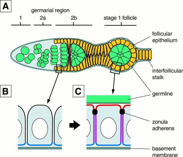
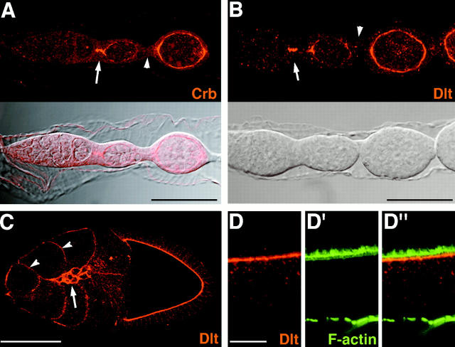
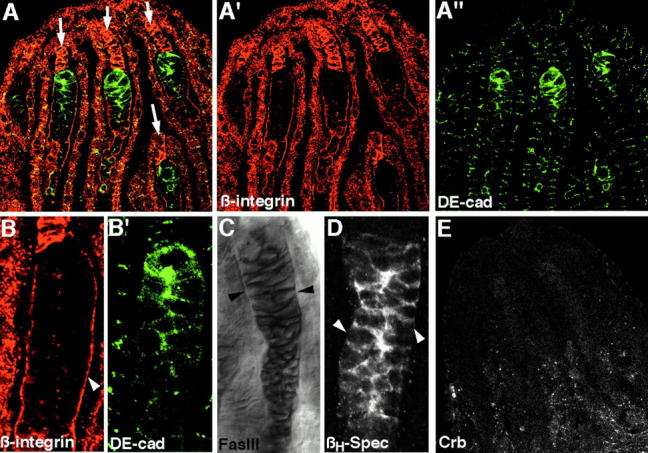
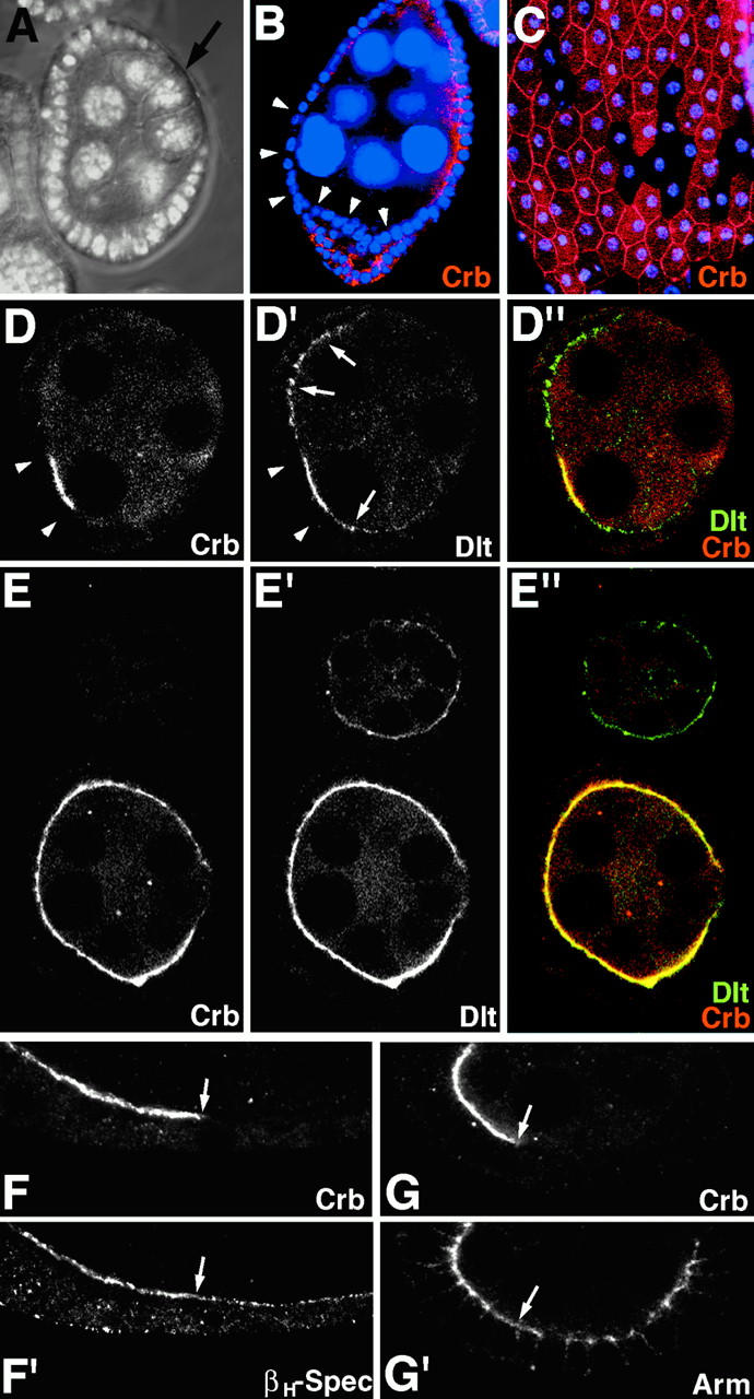
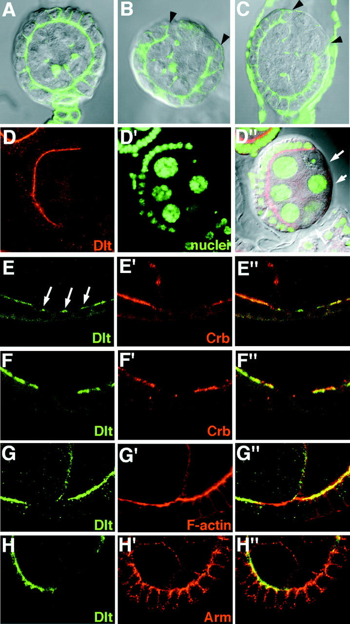
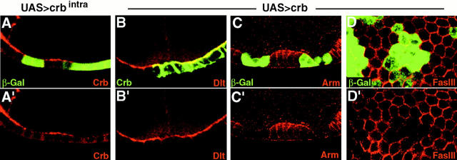
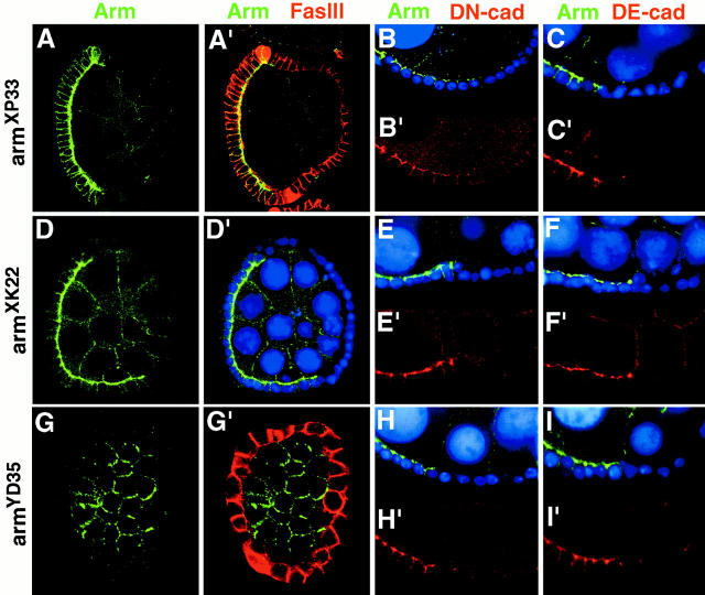
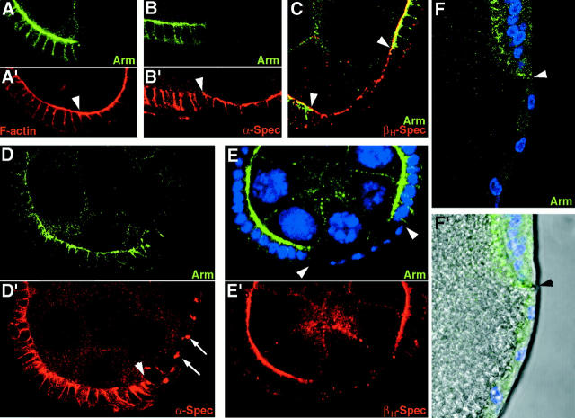
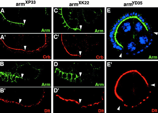
Analysis of the mechanisms that control epithelial polarization has revealed that cues for polarization are mediated by transmembrane proteins that operate at the apical, lateral, or basal surface of epithelial cells. Whereas for any given epithelial cell type only one or two polarization systems have been identified to date, we report here that the follicular epithelium in Drosophila ovaries uses three different polarization mechanisms, each operating at one of the three main epithelial surface domains. The follicular epithelium arises through a mesenchymal-epithelial transition. Contact with the basement membrane provides an initial polarization cue that leads to the formation of a basal membrane domain. Moreover, we use mosaic analysis to show that Crumbs (Crb) is required for the formation and maintenance of the follicular epithelium. Crb localizes to the apical membrane of follicle cells that is in contact with germline cells. Contact to the germline is required for the accumulation of Crb in follicle cells. Discs Lost (Dlt), a cytoplasmic PDZ domain protein that was shown to interact with the cytoplasmic tail of Crb, overlaps precisely in its distribution with Crb, as shown by immunoelectron microscopy. Crb localization depends on Dlt, whereas Dlt uses Crb-dependent and -independent mechanisms for apical targeting. Finally, we show that the cadherin-catenin complex is not required for the formation of the follicular epithelium, but only for its maintenance. Loss of cadherin-based adherens junctions caused by armadillo (beta-catenin) mutations results in a disruption of the lateral spectrin and actin cytoskeleton. Also Crb and the apical spectrin cytoskeleton are lost from armadillo mutant follicle cells. Together with previous data showing that Crb is required for the formation of a zonula adherens, these findings indicate a mutual dependency of apical and lateral polarization mechanisms.


Figures

Figure 1
Figure 1
Schematic of the formation of the FE. (A) A germarium and a stage 1 follicle. Follicle cells originate from two stem cells that are located at the transition of regions 2a and 2b (Margolis and Spradling 1995). Follicle cells surround a cluster of 16 germline cells in region 2b. Follicle cells that contact the germline cells fully polarize and form the FE, whereas a second group of follicle cells forms the interfollicular stalk by cell intercalation. (B) A follicle cell before contact to the germline. The cell is in contact to the basement membrane and has established a basal membrane (blue), whereas apical and lateral membranes have not been clearly defined. (C) A follicle cell after contact to the germline has been established. Apical (red) and lateral (purple) membranes have been formed and a zonula adherens has differentiated.
Figure 2
Figure 2
Expression of Crb and Dlt in wild-type ovaries. (A) Crb is first detected in follicle cells located between region 2b and the stage 1 follicle that will form the interfollicular stalk (arrow) and in the FE of the stage 1 follicle. Note that interfollicular stalk cells lose Crb rapidly (arrowhead), but that Crb expression is maintained in the FE, where it associates with the apical membrane. (B) Dlt shows the same distribution as Crb in early stages of oogenesis. (C) Expression of Dlt at stage 9 of oogenesis. Dlt is expressed in the FE and the border cells (arrow). In germline cells, Dlt is found at a contact site between nurse cells (arrowheads). (D) Dlt is found at the apical surface of follicle cells, but is absent from the oocyte membrane that is associated with large amounts of F-actin (D′). (D″) A merged image of D and D′. By contrast, Crb strongly accumulates in the oocyte membrane, as was shown previously (Niewiadomska et al. 1999).
Figure 3
Figure 3
IEM of Dlt in the FE and in embryonic ectoderm and epidermis. (A) Schematic of the apicolateral region of an epithelial cell after the junctional complex composed of a ZA and a septate junction has formed. The marginal zone of the apical membrane domain is an area of cell–cell contact that is found apical to the ZA (Tepass 1996). (B and C) Dlt is found throughout the apical membrane of follicle cells, but is concentrated just apical to the ZA (between arrowheads) in the marginal zone. The higher concentration of Dlt in the marginal zone is particularly apparent when the signal intensity is low as in C, where the signal is confined to the marginal zone. (D, E, and G) Examples of Dlt (D and E) and Crb (G) immunolabeling in ectodermal cells of stage 11 embryos. Labeling for Dlt and Crb are confined to the apical surface and concentrated at the marginal zone immediately apical to the ZA (between arrowheads). (F) Dlt is found apical to the ZA (arrowheads) in stage 16 embryos after the septate junction has formed (arrows). Bars, 100 μm.
Figure 4
Figure 4
Follicle cells in agametic ovaries show partial polarization. Distribution of epithelial polarity markers in agametic ovaries of flies derived from oskar301 homozygous mutant females (Lehmann and Nusslein-Volhard 1986). (A) The anterior half of a oskar301 mutant ovary double labeled for βPS-integrin (red) and DE-cadherin (green). The arrows point to individual ovarioles in which the intense DE-cadherin labeling highlights the follicle cells. (B) As in wild type (data not shown), βPS-integrin localizes to the basal membrane of follicle cells in agametic ovaries (arrowhead), whereas DE-cadherin is largely excluded from the basal membrane and accumulates in the remaining cell surface. (C) Fasciclin III is excluded from the basal membrane (arrowheads). (D) The apical marker βHeavy-spectrin is excluded from the basal membrane (arrowheads) as well. Apical and lateral markers show an overlapping distribution and concentrate at the center of the follicle cell column at the cell pole that opposes the basal cell surface. (E) An anterior half of an oskar301 mutant ovary (compare with A) labeled for Crb. Crb is not expressed in the follicle cells of oskar301 mutant ovaries.
Figure 5
Figure 5
Crb is required for the formation and maintenance of the FE. (A and B) crb11A22 mutant follicle cell clones induced before the formation of the FE may lead to epithelial discontinuities (A, arrow), or multilayering defects in posterior follicle cells (B, arrowheads indicate crb mutant cells). (C) Small crb11A22 mutant cell clones, induced after the FE has formed, show no morphological defects. A nuclear counterstain has been used in A–C. (D) Follicle with mostly crb11A22 mutant follicle cells and some crb positive cells (between arrowheads). Note the large gap in the FE between 12 and 4 o'clock. The remaining crb mutant cells, which have formed an epithelial layer retain apical Dlt (arrows), which, however, is reduced in concentration as compared with wild-type cells. (E) Wild-type follicle (bottom) and a follicle in which all cells of the FE are crb mutant (top). Small amounts of Dlt have been retained at the apical membrane of the crb mutant follicle cells. (F) crb11A22mutant clone shows reduced concentration of βHeavy-spectrin associated with the apical membrane. (G) The distribution and levels of Arm appear normal in crb mutant follicle cells. Arrows in F and G point to the boundary between mutant and wild-type cells.
Figure 6
Figure 6
Dlt is required for the formation of the FE and apical localization of Crb. (A) Wild-type stage 2 follicle stained with phalloidin. Note the prominent accumulation of F-actin at the apical surface of the cells of the FE. (B) Stage 2 and (C) stage 4 follicles containing dltdre1 mutant early clones. The FE shows gaps (between arrowheads) into which germline cells have penetrated. (D) dltMY10 mutant follicle cells, induced before the FE forms, do not form a FE, resulting in follicles with gaps in the FE (arrows). (E–H) Late dlt mutant clones. (E) dltdre1and (F) dltMY10mutant follicle cells have lost Crb. Dlt forms a central cap in the apical membrane in dltdre1 mutant cells (E, arrows). (G and H) dltMY10 mutant follicle cell clones show normal distribution of F-actin (G) and Arm (H).
Figure 7
Figure 7
Overexpression of Crb disrupts lateral markers but has little effect on Dlt distribution. (A) Follicle cells overexpressing UAS>crbintra (β-Gal) do not contain detectable amounts of endogenous Crb as detected by mAbCq4, which recognizes the extracellular part of Crb that is missing in Crbintra. (B) In follicle cells that overexpress full-length Crb (UAS>crb), Crb localizes to the apical and lateral membranes, but does not cause a substantial mislocalization of Dlt. (C and D) Levels of Arm (C) and Fasciclin III (D) are strongly reduced in some follicle cells that overexpress Crb (UAS>crb). Clones are detected by anti–β-Gal staining.
Figure 8
Figure 8
Disruption of the cadherin–catenin complex by arm mutations does not interfere with the formation of the FE. Large follicle cell clones mutant for intermediate (armXP33; A–C), strong (armXK22; D–F), and null (armYD35; G–I) alleles of arm form a FE. arm mutant cells are indicated by the absence of Arm immunoreactivity, and highlighted by Fasciclin III staining (FasIII; A′, G′) or by a nuclear stain (blue). Arm mutant cells do not express detectable amounts of DN-cadherin (DN-cad; B, E, and H) or DE-cadherin (DE-cad; C, F, and I).
Figure 9
Figure 9
Distribution of F-actin, α-Spectrin (α-Spec) and βHeavy-Spectrin (βH-Spec) in arm mutant follicle cells. (A–C) armXP33 mutant follicle cells (arrowheads point to clone boundary) show a strong reduction at the lateral membrane and an apical accumulation of F-actin (A) and α-Spectrin (B). Normal amounts of βHeavy-Spectrin are associated with the apical membrane in armXP33 mutant cells (C). (D and E) Follicle cells mutant for armXK22 (arrowheads) have a flat cell shape and show a reduction in the size of the lateral membrane domain as indicated by the α-Spectrin staining (arrows). Apical βHeavy-Spectrin is lost from armXK22 mutant follicle cells (E). (F) armYD35 mutant follicle cells (below arrowhead) show squamous cell morphology in this stage 10 follicle. Nuclei are stained blue in E and F.
Figure 10
Figure 10
Crb and Dlt distribution in arm mutant follicle cells. (A and B) armXP33 mutant follicle cells show a normal apical distribution of Crb and Dlt. (C–E) Crb is lost in armXK22 and armYD35 mutant follicle cells (C), but Dlt is retained at the apical membrane in these cells (D and E). Nuclei are stained blue in E. Clone boundaries are marked by arrowheads.

References

    1. Baumgartner S., Littleton J.T., Broadie K., Bhat M.A., Harbecke R., Lengyel J.A., Chiquet-Ehrismann R., Prokop A., Bellen H.J. A Drosophila neurexin is required for septate junction and blood-nerve barrier formation and function. Cell. 1996;87:1059–1068. - PubMed
    1. Bhat M.A., Izaddoost S., Lu Y., Cho K.O., Choi K.W., Bellen H. Discs Lost, a novel multi-PDZ domain protein, establishes and maintains epithelial polarity. Cell. 1999;96:633–645. - PubMed
    1. Bilder D., Li M., Perrimon N. Cooperative regulation of cell polarity and growth by Drosophila tumor suppressors. Science. 2000;289:113–116. - PubMed
    1. Brook W.J., Ostafichuk L.M., Piorecky J., Wilkinson M.D., Hodgetts D.J., Russell M.A. Gene expression during imaginal disc regeneration detected using enhancer-sensitive P-elements. Development (Camb.). 1993;117:1287–1297. - PubMed
    1. Byers T.J., Dubreuil R., Branton D., Kiehart D.P., Goldstein L.S. Drosophila spectrin. II. Conserved features of the α-subunit are revealed by analysis of cDNA clones and fusion proteins. J. Cell Biol. 1987;105:2103–2110. - PMC - PubMed

Publication types

MeSH terms