. 2000 Nov 13;151(4):919-30.
doi: 10.1083/jcb.151.4.919.

The subcellular localization of an aquaporin-2 tetramer depends on the stoichiometry of phosphorylated and nonphosphorylated monomers

Affiliations

The subcellular localization of an aquaporin-2 tetramer depends on the stoichiometry of phosphorylated and nonphosphorylated monomers

E J Kamsteeg et al. J Cell Biol. .

Abstract

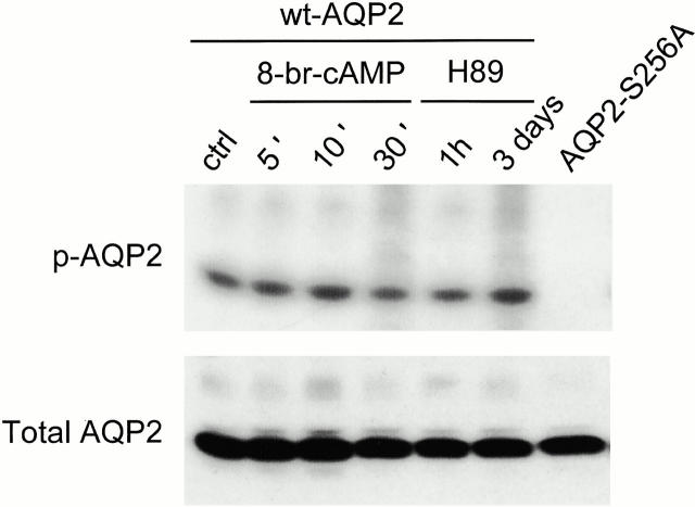
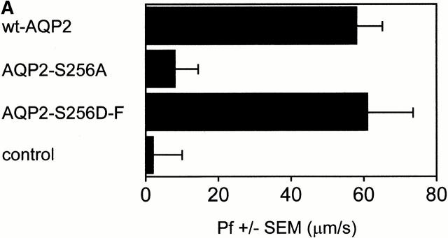
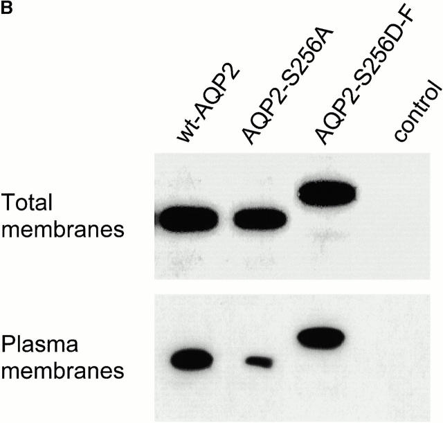
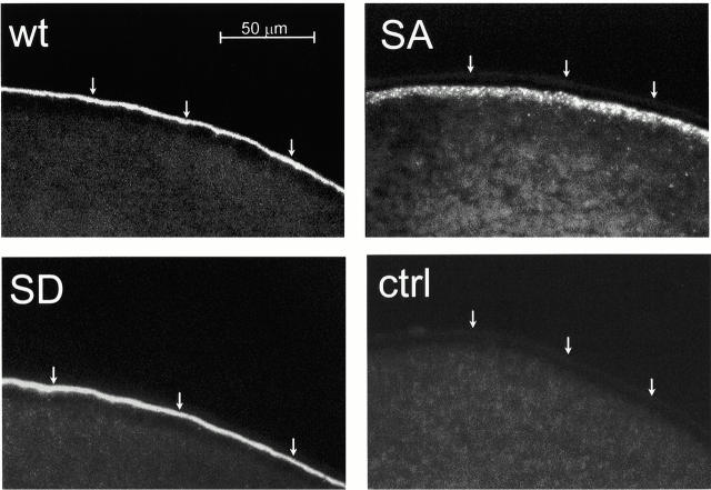
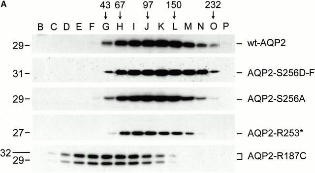
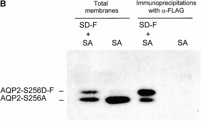
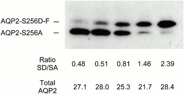
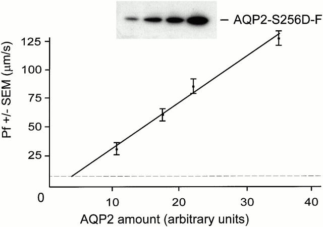
In renal principal cells, vasopressin regulates the shuttling of the aquaporin (AQP)2 water channel between intracellular vesicles and the apical plasma membrane. Vasopressin-induced phosphorylation of AQP2 at serine 256 (S256) by protein kinase A (PKA) is essential for its localization in the membrane. However, phosphorylated AQP2 (p-AQP2) has also been detected in intracellular vesicles of noninduced principal cells. As AQP2 is expressed as homotetramers, we hypothesized that the number of p-AQP2 monomers in a tetramer might be critical for the its steady state distribution. Expressed in oocytes, AQP2-S256D and AQP2-S256A mimicked p-AQP2 and non-p-AQP2, respectively, as routing and function of AQP2-S256D and wild-type AQP2 (wt-AQP2) were identical, whereas AQP2-S256A was retained intracellularly. In coinjection experiments, AQP2-S256A and AQP2-S256D formed heterotetramers. Coinjection of different ratios of AQP2-S256A and AQP2-S256D cRNAs revealed that minimally three AQP2-S256D monomers in an AQP2 tetramer were essential for its plasma membrane localization. Therefore, our results suggest that in principal cells, minimally three monomers per AQP2 tetramer have to be phosphorylated for its steady state localization in the apical membrane. As other multisubunit channels are also regulated by phosphorylation, it is anticipated that the stoichiometry of their phosphorylated and nonphosphorylated subunits may fine-tune the activity or subcellular localization of these complexes.


Figures

Figure 1
Figure 1
Phosphorylation of AQP2 in oocytes. Oocytes, injected with wt-AQP2 or AQP2-S256A cRNA, were labeled with [32P]orthophosphate for 2 d. Subsequently, oocytes were not stimulated (ctrl, AQP2-S256A), or were incubated with 5 × 10⁻⁴ M 8-Br-cAMP or 10−4 M H89 for the indicated time periods. Total membranes were isolated and divided into two portions. One portion was solubilized, AQP2 was immunoprecipitated and subjected to PAGE, the gel was dried, and labeled AQP2 (p-AQP2) was visualized using autoradiography. The other portion was directly immunoblotted for AQP2 (Total AQP2).
Figure 2
Figure 2
Osmotic water permeability (Pf) and immunoblot analysis of oocytes expressing wild-type or mutant AQP2. (A) Oocytes were not injected (control), or were injected with cRNA encoding wt-AQP2, AQP2-S256A, or AQP2-S256D-F. 2 d after injection, the mean Pfs ± SEM (in μm/s) of at least 12 oocytes were determined in a standard swelling assay. (B) From the oocytes that were used for the Pf measurements, total membranes and plasma membranes were isolated and immunoblotted for AQP2.
Figure 2
Figure 2
Osmotic water permeability (Pf) and immunoblot analysis of oocytes expressing wild-type or mutant AQP2. (A) Oocytes were not injected (control), or were injected with cRNA encoding wt-AQP2, AQP2-S256A, or AQP2-S256D-F. 2 d after injection, the mean Pfs ± SEM (in μm/s) of at least 12 oocytes were determined in a standard swelling assay. (B) From the oocytes that were used for the Pf measurements, total membranes and plasma membranes were isolated and immunoblotted for AQP2.
Figure 3
Figure 3
Immunocytochemistry of oocytes expressing wild-type or mutant AQP2 proteins. 2 d after injection, oocytes injected with cRNA encoding wt-AQP2 (wt), AQP2-S256A (SA), or AQP2-S256D-F (SD), and noninjected oocytes (ctrl) were fixed and embedded in paraffin. In 5-μm sections, AQP2 was visualized using rabbit AQP2 antibodies followed by Alexa-594–conjugated anti–rabbit IgG. Arrows indicate the plasma membrane.
Figure 4
Figure 4
Oligomerization state of AQP2 proteins and coimmunoprecipitation of AQP2-S256A with AQP2-S256D-F. (A) Oocytes were injected with cRNAs encoding wt-AQP2, AQP2-S256A, AQP2-S256D-F, AQP2-R253*, or AQP2-R187C. 3 d after injection, membranes of these oocytes were solubilized in 4% deoxycholate and subjected to sucrose gradient sedimentation centrifugation. Fractions were collected from the top, of which fractions B–P were immunoblotted for AQP2. The fractions with peak intensities of the marker proteins ovalbumin, BSA, phosphorylase B, yeast alcohol dehydrogenase, and catalase are indicated by their molecular masses (45, 67, 97, 150, and 232, respectively). The molecular masses (in kD) of the AQP2 proteins are indicated on the left. (B) Membranes of oocytes expressing AQP2-S256A alone (SA) or in combination with AQP2-S256D-F (SD-F + SA) were directly subjected to immunoblotting for AQP2 (Total membranes) or first solubilized in deoxycholate, immunoprecipitated with FLAG antibodies, and then subjected to immunoblotting (Immunoprecipitations with α-FLAG). Due to the FLAG tag, the molecular mass of AQP2-S256D-F is higher than that of AQP2-S256A.
Figure 4
Figure 4
Oligomerization state of AQP2 proteins and coimmunoprecipitation of AQP2-S256A with AQP2-S256D-F. (A) Oocytes were injected with cRNAs encoding wt-AQP2, AQP2-S256A, AQP2-S256D-F, AQP2-R253*, or AQP2-R187C. 3 d after injection, membranes of these oocytes were solubilized in 4% deoxycholate and subjected to sucrose gradient sedimentation centrifugation. Fractions were collected from the top, of which fractions B–P were immunoblotted for AQP2. The fractions with peak intensities of the marker proteins ovalbumin, BSA, phosphorylase B, yeast alcohol dehydrogenase, and catalase are indicated by their molecular masses (45, 67, 97, 150, and 232, respectively). The molecular masses (in kD) of the AQP2 proteins are indicated on the left. (B) Membranes of oocytes expressing AQP2-S256A alone (SA) or in combination with AQP2-S256D-F (SD-F + SA) were directly subjected to immunoblotting for AQP2 (Total membranes) or first solubilized in deoxycholate, immunoprecipitated with FLAG antibodies, and then subjected to immunoblotting (Immunoprecipitations with α-FLAG). Due to the FLAG tag, the molecular mass of AQP2-S256D-F is higher than that of AQP2-S256A.
Figure 5
Figure 5
Semiquantification of the amounts and ratios of AQP2-S256A and AQP2-S256D-F in coinjections. Copy RNAs encoding AQP2-S256D-F and AQP2-S256A were mixed in the ratios 1:3, 1:2, 1:1, 2:1, and 3:1 (from left to right) and a total of 0. 4 ng was injected into oocytes. 2 d later, the Pf values were determined (see Table ) and total membranes were isolated and immunoblotted for AQP2. The individual signals of AQP2-S256A (SA) and AQP2-S256D-F (SD) were scanned and the amounts of expressed protein were semiquantified (in arbitrary units) using the scanned signals of a twofold dilution series of wt-AQP2 as a standard. From these data, the SD/SA ratio and total AQP2 amounts were calculated.
Figure 6
Figure 6
The relation between the expressed amounts and conferred water permeabilities of AQP2-S256D-F. Of oocytes, injected with 0.1, 0.2, 0.3, or 0.4 ng of AQP2-S256D-F cRNA, the water permeability (Pf ± SEM in μm/s) was measured. Total membranes were isolated and immunoblotted for AQP2 (AQP2-S256D-F; insert). The signals were scanned and the amounts of expressed AQP2-S256D-F (AQP2 amount in arbitrary units) were semiquantified as described in the legend to Fig. 5. The linear relation between the amount of AQP2-S256D-F protein and the conferred water permeability was: Pf = (3.97 × amount of AQP2 protein expected in membrane) − 9. 7. For expression levels lower than obtained for 0.1 ng AQP2-S256D-F cRNA, the relation was extrapolated to the Pf level of control oocytes (8 ± 4 μm/s, dotted line).

References

    1. Bao J., Alroy I., Waterman H., Schejter E.D., Brodie C., Gruenberg J., Yarden Y. Threonine phosphorylation diverts internalized epidermal growth factor receptors from a degradative pathway to the recycling endosome. J. Biol. Chem. 2000;275:26178–26186. - PubMed
    1. Brown D. Membrane recycling and epithelial cell function. Am. J. Physiol. 1989;256:F1–F12. - PubMed
    1. Brown D., Stow J.L. Protein trafficking and polarity in kidney epitheliumfrom cell biology to physiology. Physiol. Rev. 1996;76:245–297. - PubMed
    1. Brown D., Weyer P., Orci L. Vasopressin stimulates endocytosis in kidney collecting duct principal cells. Eur. J. Cell Biol. 1988;46:336–341. - PubMed
    1. Cheng C., Prince L.S., Snyder P.M., Welsh M.J. Assembly of the epithelial Na+ channel evaluated using sucrose gradient sedimentation analysis. J. Biol. Chem. 1998;273:22693–22700. - PubMed

Publication types

MeSH terms