. 2000 Nov 13;151(4):931-44.
doi: 10.1083/jcb.151.4.931.

Connexin43 deficiency causes delayed ossification, craniofacial abnormalities, and osteoblast dysfunction

Affiliations

Connexin43 deficiency causes delayed ossification, craniofacial abnormalities, and osteoblast dysfunction

F Lecanda et al. J Cell Biol. .

Abstract

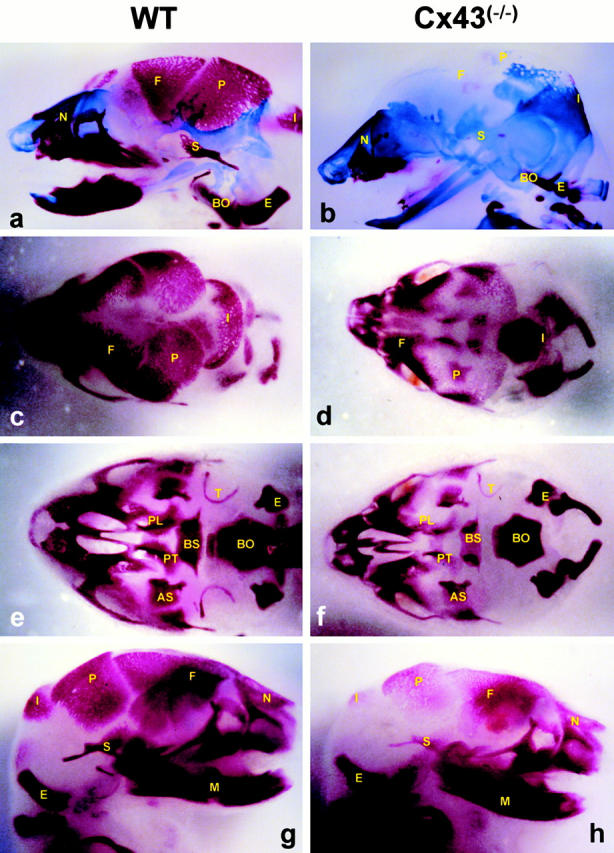
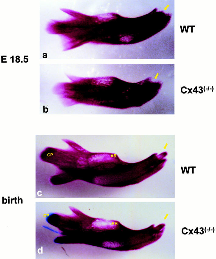
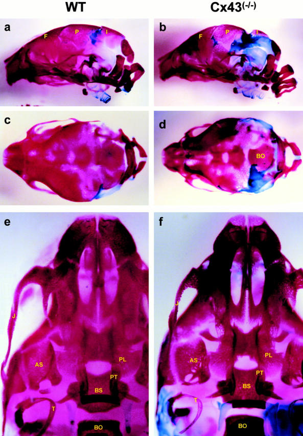
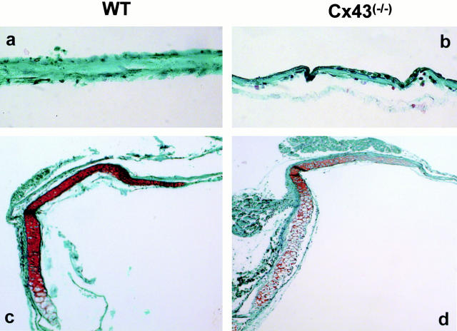
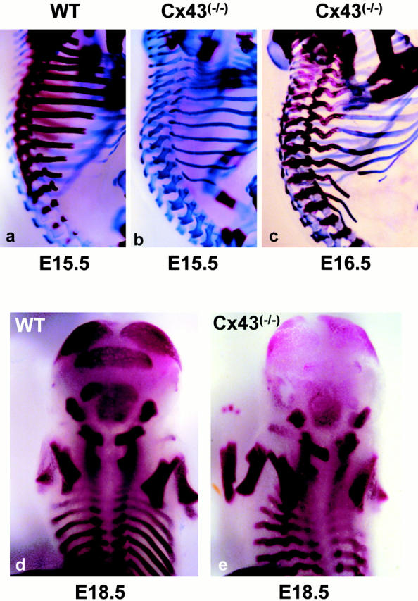
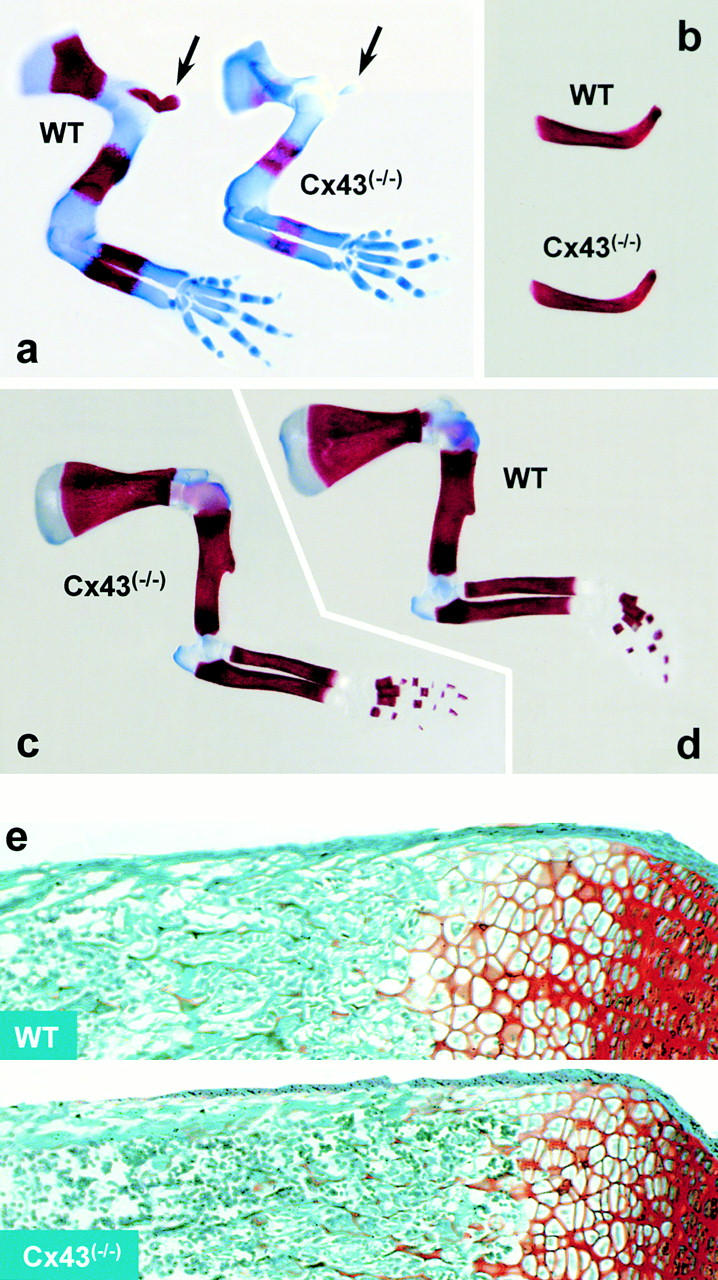
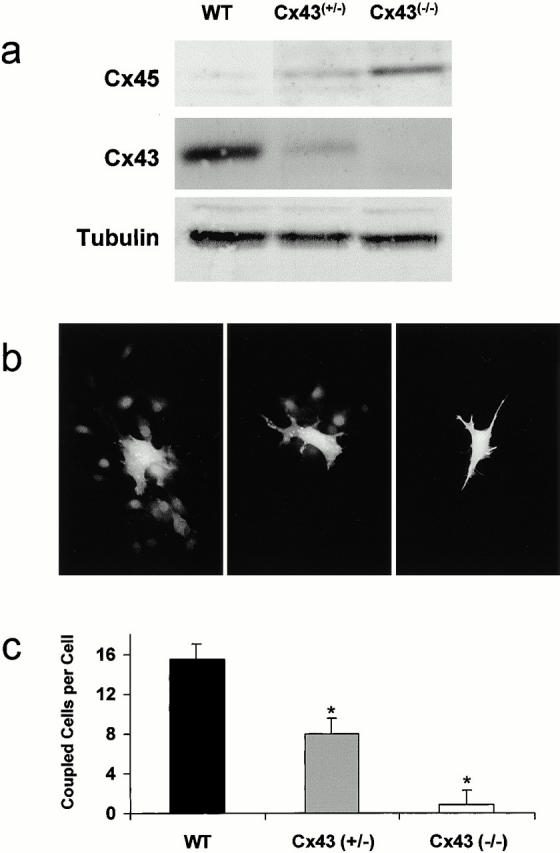
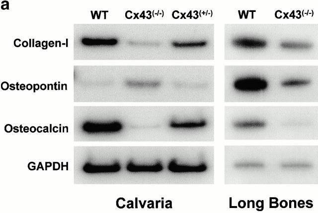
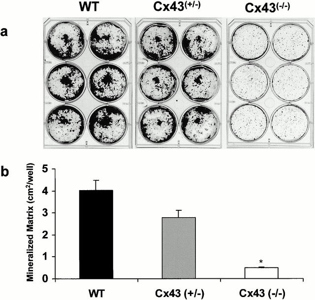
Connexin(Cx)43 is the major gap junction protein present in osteoblasts. We have shown that overexpression of Cx45 in osteoblasts expressing endogenous Cx43 leads to decreased cell-cell communication (Koval, M., S.T. Geist, E.M. Westphale, A.E. Kemendy, R. Civitelli, E.C. Beyer, and T.H. Steinberg. 1995. J. Cell Biol. 130:987-995) and transcriptional downregulation of several osteoblastic differentiation markers (Lecanda, F., D.A. Towler, K. Ziambaras, S.-L. Cheng, M. Koval, T.H. Steinberg, and R. Civitelli. 1998. Mol. Biol. Cell 9:2249-2258). Here, using the Cx43-null mouse model, we determined whether genetic deficiency of Cx43 affects skeletal development in vivo. Both intramembranous and endochondral ossification of the cranial vault were delayed in the mutant embryos, and cranial bones originating from migratory neural crest cells were also hypoplastic, leaving an open foramen at birth. Cx43-deficient animals also exhibited retarded ossification of the clavicles, ribs, vertebrae, and limbs, demonstrating that skeletal abnormalities are not restricted to a neural crest defect. However, the axial and appendicular skeleton of Cx43-null animals were essentially normal at birth. Cell to cell diffusion of calcein was poor among Cx43-deficient osteoblasts, whose differentiated phenotypic profile and mineralization potential were greatly impaired, compared with wild-type cells. Therefore, in addition to the reported neural crest cell defect, lack of Cx43 also causes a generalized osteoblast dysfunction, leading to delayed mineralization and skull abnormalities. Cell to cell signaling, mediated by Cx43 gap junctions, was critical for normal osteogenesis, craniofacial development, and osteoblastic function.


Figures

Figure 1
Figure 1
Skull development in Cx43−/− embryos. (a and b) Lateral view of the skull of alizarin red/alcian blue–stained E16.5 wild-type (WT) and Cx43−/− embryos. (c and d) Dorsal, (e and f) ventral, and (g and h) lateral views of the skull of alizarin red–stained E18.5 (c, e, and g) wild-type and (d, f, and h) Cx43−/− embryos. (g and h) Lateral view on the skull at E18.5 shows the incipient mineralization of parietal, nasal, frontal, and squamous parts of the temporal bones in Cx43-null mice, though all these elements remain developmentally hypomineralized, especially around their rostral boundaries. F, frontal bone; P, parietal bone; S, squamous part of the temporal bone; I, interparietal bone; E, exoccipital bone; M, mandible; T, tympanic ring; PL, palatine bone; PT, pterygoid bone; BS, basisphenoid bone; AS, alisphnoid bone; and BO, basioccipital bone.
Figure 2
Figure 2
Mandible development in Cx43−/− mice. Alizarin red/alcian blue staining of the mandibles at (a and b) E18.5 and at (c and d) birth of wild-type (WT) and Cx43−/− littermates. In newborn mutants, the mandible was smaller, with a more rounded alveolar ridge and a flatter arch. This malformation contributes to the slightly smaller and more pointed snout in mutant animals. In some Cx43-null newborn mice, cartilaginous primordia of the temporal bone and the condylar region of the mandible were still present at (d) birth Note, (b, arrow) the lack of the incisor tooth at E18.5 in the Cx43−/− embryo and (d) the less prominent tooth at birth. Also, note the less pronounced alveolar ridge (AR) in the mutant animals and the cartilaginous coronoid process (CP).
Figure 3
Figure 3
Skull development in Cx43−/− at birth. (a and b) Lateral and (c and d) dorsal views of the cranial vault at birth of (a and c) wild-type and (b and d) Cx43−/− littermates at birth. Note the prominent cartilagenous primordia of the occipital bone in the homozygous mutants, whereas the parietal, interparietal, and supraoccipital bones are thinner and less developed, allowing a direct view of the cranial base. A ventral view of the cranial base of (e) wild-type and (f) Cx43-null littermates at birth. The horizontal laminae of the palatine bones were slightly smaller, and the alisphenoid was misshapen, leaving a larger unmineralized area delimited by the palatine, alisphenoid, and temporal bones. The horizontal portion of the palatine bones and the palatine processes of the maxillar bone, collectively referred to as palatal shelves, were present. The tympanic ring was reduced in size and thinner. The jugal bone of the zygomatic arch was shorter, resulting in a misshapen zygomatic arch. Overall, the nasal and maxillary bones were slightly smaller, resulting in a more pointed and smaller snout in the Cx43-null animals. J, jugal part of the zygomatic bone; F, frontal bone; P, parietal bone; S, squamous part of the temporal bone; I, interparietal bone; E, exoccipital bone; M, mandible; T, tympanic ring; PL, palatine bone; PT, pterygoid bone; BS, basisphenoid bone; AS, alisphenoid bone; and BO, basioccipital bone.
Figure 4
Figure 4
Sagittal sections of the (a and b) parietal and (c and d) occipital bones of the (a and c) wild-type and (b and d) Cx43−/− mice stained with safranin/fast green. Note that the parietal bone is thinner and more brittle in the Cx43 homozygous mutants. (d) The cartilaginous anlage of the occipital bone is more prominent in the Cx43−/− animals.
Figure 5
Figure 5
Axial development in Cx43−/− embryos. Lateral view of the thoracic vertebrae stained with alizarin red/alcian blue of (a and b) E15.5 and (c) E16.5 (a and d) wild-type embryos and (b, c, and e) Cx43−/− homozygous mutants. (b and c) Note the delayed ossification of the vertebrae and ribs and the thinner and deformed ribs in the mutant animals. Posterior view of the head and thorax of alizarin red–stained (d) wild-type and (e) Cx43−/− embryos at E18.5 showing a similar ossification of the vertebrae, limbs, and exoccipital bones, but delayed ossification of the parietal, interparietal, and supraoccipital bones. Note that the two ossification centers of the supraoccipital bone have not fused in the Cx43−/−.
Figure 6
Figure 6
Development of the appendicular skeleton in Cx43−/− embryos. Front limbs at (a) E14.5, (b) clavicle at birth, and (c and d) front limbs at birth were stained with alizarin red/alcian blue. Note, the delayed mineralization of the scapula and the long limb bones (humerus, radius, and cubitus) in the mutant embryos at (a) E14.5 compared with (c and d) normal birth. Also, note the lack of both intramembranous and endochondral ossification of the clavicle, present only as a small cartilaginous template in the (a, arrows) Cx43−/− animals at E14.5. However, the clavicles appeared normal at (b) birth. (e) Longitudinal sections of safranin O/fast green–stained femurs of wild-type and Cx43−/− neonates. The size and morphology of the growth plate is apparently normal, as is the primary spongiosa in the homozygous mutant bone.
Figure 7
Figure 7
Cx expression and cell coupling in calvaria osteoblasts derived from wild-type (WT), heterozygous Cx43+/−, and homozygous Cx43−/− mice at birth. (a) Immunoblot of whole cell lysate using antibodies against Cx45 and Cx43. Tubulin was used to control for protein loading in each lane. (b) Diffusion of calcein among calvaria osteoblasts of the different genotype groups. Dye coupling was assessed two hours after “parachuting” calcein-loaded donor cells onto an unlabeled monolayer and by counting neighboring cells that have taken the fluorescent dye in 20 microscopic fields in each cell preparation. Cells of the same genotypic group were used as donor cells after calcein loading. (c) Quantitation of dye coupling in each genotype group. The number of cells taking calcein from a single preloaded donor cell was counted as a measure of dye coupling. Data represent the average ± SD of eight random microscopic fields in 13-mm coverslips (ten coverslips per isolate, pooled data from four cell isolates per genotype group). *P < 0.05, Mann-Whitney U test.
Figure 8
Figure 8
Production of bone matrix proteins by osteoblasts derived from either calvaria or long bones (femur and tibia) of newborn animals. (a) Total RNA was isolated from cultures of wild-type, heterozygous Cx43+/−, and homozygous Cx43−/− cells, after they reached confluency, as indicated. Steady state mRNA levels of type I collagen (Collagen-I), osteopontin, osteocalcin, and glyceraldehyde phosphate dehydrogenase were assessed by semiquantitave RT-PCR. (b) Western analysis of type I collagen (Collagen-I), osteopontin, and tubulin in the three genotype groups using calvaria cells.
Figure 8
Figure 8
Production of bone matrix proteins by osteoblasts derived from either calvaria or long bones (femur and tibia) of newborn animals. (a) Total RNA was isolated from cultures of wild-type, heterozygous Cx43+/−, and homozygous Cx43−/− cells, after they reached confluency, as indicated. Steady state mRNA levels of type I collagen (Collagen-I), osteopontin, osteocalcin, and glyceraldehyde phosphate dehydrogenase were assessed by semiquantitave RT-PCR. (b) Western analysis of type I collagen (Collagen-I), osteopontin, and tubulin in the three genotype groups using calvaria cells.
Figure 9
Figure 9
In vitro mineral deposition by calvaria osteoblasts. Cells isolated from calvaria of newborn wild-type (WT), heterozygous Cx43+/−, and homozygous Cx43−/− mice were grown in mineralizing medium, containing β-glycerophosphate and ascorbic acid, for three weeks. (a) Cultures were stained with Von Kossa. (b) The extent of mineral deposited in the extracellular matrix was quantitated as the surface area (in cm2) covered by black stain in each well (9.6 cm2) using a computerized image analyzer. The illustration and bar chart are representative of identical experiments performed in four independent cell isolates per genotype group.

References

    1. Becker D.L., McGonnell I., Makarenkova H.P., Patel K., Tickle C., Lorimer J., Green C.R. Roles for alpha 1 connexin in morphogenesis of chick embryos revealed using a novel antisense approach. Dev. Genet. 1999;24:33–42. - PubMed
    1. Bradford M. A rapid and sensitive method for the quantitation of microgram quantities of protein utilizing the principle of protein dye binding. Anal. Biochem. 1976;72:248–254. - PubMed
    1. Chen H., Ovchinnikov D., Pressman C.L., Aulehla A., Lun Y., Johnson R.L. Multiple calvarial defects in lmx1b mutant mice. Dev. Genet. 1998;22:314–320. - PubMed
    1. Chen Y., Bei M., Woo I., Satokata I., Maas R. Msx1 controls inductive signaling in mammalian tooth morphogenesis. Development. 1996;122:3035–3044. - PubMed
    1. Cheng S.-L., Yang J.W., Rifas L., Zhang S.-F., Avioli L.V. Differentiation of human bone marrow osteogenic stromal cells in vitroinduction of the osteoblast phenotype by dexamethasone. Endocrinology. 1994;134:277–286. - PubMed

Publication types

MeSH terms