. 2000 Nov 21;97(24):13245-50.
doi: 10.1073/pnas.230442897.

Paired Ig-like receptor homologs in birds and mammals share a common ancestor with mammalian Fc receptors

Affiliations

Paired Ig-like receptor homologs in birds and mammals share a common ancestor with mammalian Fc receptors

G Dennis Jr et al. Proc Natl Acad Sci U S A. .

Abstract

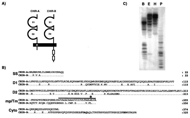
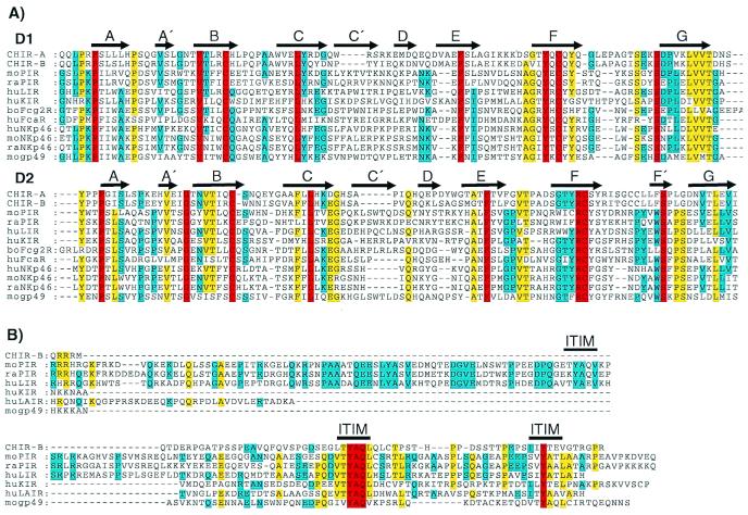
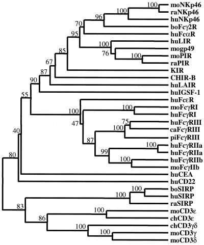
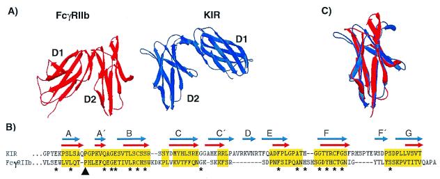
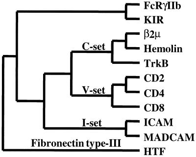
Paired Ig-like receptors (PIR) that can reciprocally modulate cellular activation have been described in mammals. In the present study, we searched expressed sequence tag databases for PIR relatives to identify chicken expressed sequence tags predictive of approximately 25% amino acid identity to mouse PIR. Rapid amplification of cDNA ends (RACE)-PCR extension of expressed sequence-tag sequences using chicken splenic cDNA as a template yielded two distinct cDNAs, the sequence analysis of which predicted protein products with related extracellular Ig-like domains. Chicken Ig-like receptor (CHIR)-A was characterized by its transmembrane segment with a positively charged histidine residue and short cytoplasmic tail, thereby identifying CHIR-A as a candidate-activating receptor. Conversely, CHIR-B was characterized by its nonpolar transmembrane segment and cytoplasmic tail with two immunoreceptor tyrosine-based inhibitory motifs, indicating that it may serve as an inhibitory receptor. The use of CHIR amino acid sequences in a search for other PIR relatives led to the recognition of mammalian Fc receptors as distantly related genes. Comparative analyses based on amino acid sequences and three-dimensional protein structures provided molecular evidence for common ancestry of the PIR and Fc receptor gene families.


Figures

Figure 1
Figure 1
(A) Schematic representation of the predicted CHIR-A and CHIR-B molecules with two Ig-like extracellular domains. CHIR-A encodes a short cytoplasmic region and a transmembrane segment with a charged histidine (H) residue. CHIR-B possesses a long cytoplasmic tail with two ITIM units (outline boxes) and a nonpolar transmembrane region. (B) Comparison of the CHIR-A and CHIR-B amino acid sequences. In this alignment, the relatives are numbered with reference to the start of the signal sequence. Conserved residues in CHIR-B are represented as dots. The solid bar indicates the putative transmembrane region; an asterisk marks the positively charged histidine residue of CHIR-A; and the ITIM-units of CHIR-B are highlighted in bold. (C) Southern blot analysis of the Chir gene family. DNA from nucleated chicken erythrocytes was digested with BamHI (B), EcoRI (E), HindIII (H), and PstI (P) and analyzed with a probe corresponding to the extracellular region of CHIR-B. Accession nos: CHIR-A, AF306851; CHIR-B, AF306852.
Figure 2
Figure 2
(A) Sequence comparison of the two amino-terminal Ig-like domains of CHIR-A, CHIR-B, mouse (mo) PIR-B (AF038149), moNKp46 (AJ223765), mogp49-B (2997305), rat (ra) PIR-B (AF16936), raNKp46 (AF082533), human (hu) LIR-1 (AF009220), huKIR2DL1 (AF022049), huFcαR (U4774), huNKp46 (AJ001383), and bovine (bo) Fcγ2R (2136749). Gaps in the alignment are indicated by dashes, and bold arrows represent regions of β-stranded secondary structures (A–G) designated according to the crystal structure of human KIR (42). Residues identical in all of the aligned sequences are in red, yellow indicates 80% of the aligned sequences are identical, and blue indicates 60% identity for the aligned sequences. (B) Comparison of the CHIR-B, moPIR-B (AF038149), mogp49-B (2997305), raPIR-B (AF16936), huLIR-1 (AF009220), huKIR (AF022049), and huLAIR (AF013249) cytoplasmic tails, with gaps in this alignment indicated by dashes. Bold lines indicate moPIR-B ITIMs demonstrated to have inhibitory function. Red indicates residues that are identical in all of the aligned sequences, yellow indicates identity in 60% of the aligned sequences, and blue indicates 40% identity for the aligned sequences.
Figure 3
Figure 3
Dendrogram of implied relationships among CHIR-like sequences identified in PSI-BLAST searches. Amino acid sequences for CHIR-B, moPIR-B (AF038149), moNKp46 (AJ223765), mogp49-B (2997305), moFcγRI (AF143180), moFcγRIIb (U31803), raPIR-B (AF16936), raNKp46 (AF082533), raSIRP (AAC18089), huLIR-1 (AF009220), huKIR2DL1 (AF022049), huFcαR (U4774), huNKp46 (AJ001383), huLAIR (AF013249), huFcɛRI (J03605), huFcγRI (L03418), huFcγRIII (AB032414), huFcγRIIa (M28697), huFcγRIIa′ (M31932), huFcγRIIb (U87564), huCD22 (S61375), huCEA (X16356), huSIRP (CAB46661), boFcγ2R (2136749), boSIRP (CAA71943), cat (ca) FcγRIII (AB025315), and pig (pi) FcγRIII (Q28942) were aligned using CLUSTALX and the Gonnet series substitution matrix. Human CD22 and carcinoembryonic antigen (CEA) were identified after the third iteration. Human, rat, and bovine SIRPs were not identified in the iterative searches but are included to provide a measure of tree topology (see text). Optimal tree topology was estimated by cluster analysis using the weighted pair-group method. Branch values represent percent bootstrap support after 500 replicates. The tree was rooted by the inclusion of mouse CD3ɛ (A31348), γ (CAA68667), and δ (CAA26198) and chicken CD3ɛ (Q98910) and γδ (A39171) as primordial Ig-like domains.
Figure 4
Figure 4
(A) Crystal structures of the extracellular Ig-like domains of FcγRIIb (red) and KIR (blue). The second domains (D2) are placed in the same orientation to illustrate the relative difference in orientation between FcγRIIb D1 and KIR D1. (B) Structure-based alignment of FcγRIIb D2 and KIR D2. Gaps are indicated by dashes, and residues used in rms deviation calculations are highlighted in yellow. The β-stranded domain topologies are indicated for FcγRIIb by red arrows and for KIR by blue arrows. The black arrowhead indicates the cis-proline residue contributing to the A to A′-strand switch, and asterisks indicate identical residues in both sequences. (C) Structural overlay of FcγRIIb (red) and KIR (blue) D2 depicting their overall similarity. Structural decorations, alignments, overlays, and rms deviation calculations were performed using Swiss-pdb Viewer (30).
Figure 5
Figure 5
Structure-based dendrogram of inferred relationships between Ig domain-containing receptors. An all-against-all structural distance matrix was determined using FcγRIIb domain (D)2 (PDB entry name 2FCBA), human KIR-D2 (1NKR), β2 μ-D1 (1BMG), Hemolin-D1 (1BIH), TrkB-D1 (1WWB), CD2-D1 (1HNF), CD4-D1 (3CD4), CD8-D1 (1CD8), ICAM-D1 (1IAM), MadCAM-D1 (1BQS), and HTF-D1 (1BOY). The optimal tree topology was estimated by parsimony analysis with global rearrangements. With the exception of FcγRIIb and KIR, domain classifications are indicated along branches according to the SCOP database. A fibronectin type III (HTF) domain serves as an outgroup to root the tree.

References

    1. Kubagawa H, Burrows P D, Cooper M D. Proc Natl Acad Sci USA. 1997;94:5261–5266. - PMC - PubMed
    1. Hayami K, Fukuta D, Nishikawa Y, Yamashita Y, Inui M, Ohyama Y, Hikida M, Ohmori H, Takai T. J Biol Chem. 1997;272:7320–7327. - PubMed
    1. Kubagawa H, Chen C C, Ho L H, Shimada T S, Gartland L, Mashburn C, Uehara T, Ravetch J V, Cooper M D. J Exp Med. 1999;189:309–317. - PMC - PubMed
    1. Yamashita Y, Ono M, Takai T. J Immunol. 1998;161:4042–4047. - PubMed
    1. Maeda A, Kurosaki M, Ono M, Kurosaki T. J Exp Med. 1998;188:991–995. - PMC - PubMed

Publication types

MeSH terms

Associated data

LinkOut - more resources