. 2000 Nov 21;97(24):13354-9.
doi: 10.1073/pnas.240347797.

Parkin functions as an E2-dependent ubiquitin- protein ligase and promotes the degradation of the synaptic vesicle-associated protein, CDCrel-1

Affiliations

Parkin functions as an E2-dependent ubiquitin- protein ligase and promotes the degradation of the synaptic vesicle-associated protein, CDCrel-1

Y Zhang et al. Proc Natl Acad Sci U S A. .

Abstract

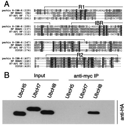
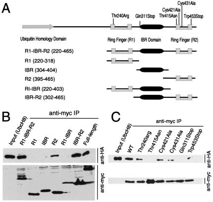
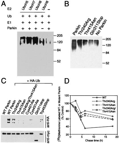
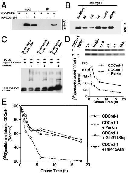
Parkinson's disease is a common neurodegenerative disorder in which familial-linked genes have provided novel insights into the pathogenesis of this disorder. Mutations in Parkin, a ring-finger-containing protein of unknown function, are implicated in the pathogenesis of autosomal recessive familial Parkinson's disease. Here, we show that Parkin binds to the E2 ubiquitin-conjugating human enzyme 8 (UbcH8) through its C-terminal ring-finger. Parkin has ubiquitin-protein ligase activity in the presence of UbcH8. Parkin also ubiquitinates itself and promotes its own degradation. We also identify and show that the synaptic vesicle-associated protein, CDCrel-1, interacts with Parkin through its ring-finger domains. Furthermore, Parkin ubiquitinates and promotes the degradation of CDCrel-1. Familial-linked mutations disrupt the ubiquitin-protein ligase function of Parkin and impair Parkin and CDCrel-1 degradation. These results suggest that Parkin functions as an E3 ubiquitin-protein ligase through its ring domains and that it may control protein levels via ubiquitination. The loss of Parkin's ubiquitin-protein ligase function in familial-linked mutations suggests that this may be the cause of familial autosomal recessive Parkinson's disease.


Figures

Figure 1
Figure 1
Parkin interacts with the E2 ubiquitin-conjugating enzyme, UbcH8. (A) Multiple sequence alignment of ring-finger domains of Parkin (BAA25751), HHARI (NP005735), H7-AP1 (CAB45870), and UIP28 (AAD24572). Conserved residues are highlighted, and the ring fingers (R1 and R2) and the IBR domain are indicated. Abbreviations: HHARI, ariadne homologue (Homo sapiens); H7-AP1, UbcH7-binding protein; UIP28, UbcM4 interacting protein 28. (B) Lysates prepared from HEK 293 cells transfected with pRK5-myc-Parkin and pRK5-HAUbcH5, pRK5-HAUbcH7, or pRK5-HAUbcH8 were subjected to immunoprecipitation (IP) with anti-myc antibody followed by anti-HA immunoblotting. This experiment was replicated three times with similar results.
Figure 2
Figure 2
UbcH8 interacts preferentially with the C-terminal R2 ring-finger domain of Parkin. (A) Schematic representation of putative functional domains of Parkin used in the mapping experiments. (B) Lysates prepared from HEK 293 cells transfected with pRK5-HA-UbcH8 and various Myc-tagged Parkin domain constructs were subjected to immunoprecipitation (IP) with anti-myc antibody followed by anti-HA immunoblotting (Upper). The blot was stripped and reprobed with the anti-myc antibody (Lower) to illustrate that equivalent levels of the Parkin constructs were present in the extracts. This experiment was replicated three times with similar results. (C) Lysates prepared from HEK 293 cells transfected with pRK5-HA-UbcH8 and various Myc-tagged Parkin mutant constructs (see text for details) were subjected to immunoprecipitation (IP) with anti-myc antibody followed by anti-HA immunoblotting (Upper). The blot was stripped and reprobed with the anti-Myc antibody (Lower) to illustrate that equivalent levels of the Parkin constructs were present in the extracts. This experiment was replicated three times with similar results.
Figure 3
Figure 3
Parkin is involved in E2-dependent self-ubiquitination both in vitro and in vivo. (A) In vitro translated Parkin (5 μl) was incubated with 100 ng of E1, 10 μg of ubiquitin (Ub) with 5 μl of bacterially expressed UbcH8, UbcH7, UbH5, or control bacteria lysate in the ubiquitination reaction buffer for 1.5 h at 30°C. The reaction products were immunoblotted with anti-ubiquitin antibody. Molecular weight markers are indicated. This experiment was replicated three times with similar results. (B) In vitro translated Parkin (5 μl) or Parkin mutants were incubated with 100 ng of E1, 10 μg of Ub with 5 μl of bacterially expressed UbcH8 in the ubiquitination reaction buffer for 1.5 h at 30°C. The reaction products were immunoblotted with anti-ubiquitin antibody. Molecular weight markers are indicated. This experiment was replicated three times with similar results. (C) Lysates prepared from HEK 293 cells transfected with pRK5-myc-Parkin or myc-tagged mutant Parkin constructs and pMT123-HA-ubiquitin were subjected to immunoprecipitation (IP) with anti-myc antibody followed by anti-HA immunoblotting (Upper). The blot was stripped and reprobed with the anti-myc antibody (Lower) to illustrate that equivalent levels of the Parkin constructs were present in the extracts. This experiment was replicated three times with similar results. (D) Familial-linked Parkin mutants impair Parkin degradation. HEK 293 cells were transfected with pRK5-myc-Parkin, pRK5-myc-ParkinThr240Arg, pRK5-myc-Parkin-Thr415Asn, and pRK5-myc-ParkinThr240Arg/Thr415Asn plasmids using Lipofectamine. 24 h after transfection, cells were washed and incubated with methionine-free medium for 1 h. The cells were pulsed-chased with 100 μCi of [35S]methionine and were harvested at the indicated times for immunoprecipitation with anti-myc antibody. The immunoprecipitates were resolved on a 10% SDS/PAGE gel and visualized and quantitated with a phosphoimager. This experiment was replicated with similar results.
Figure 4
Figure 4
Parkin interacts with, ubiquitinates and promotes the degradation of CDCrel-1. (A) Lysates prepared from HEK 293 cells transfected with pRK5-HA-CDCrel-1 and pRK5-myc-Parkin constructs were subjected to immunoprecipitation (IP) with anti-myc antibody followed by anti-HA immunoblotting. This experiment was replicated three times with similar results. (B) The ring-finger domains of Parkin interact with CDCrel-1. Lysates prepared from HEK 293 cells transfected with pRK5-HA-CDCrel-1 and myc-tagged Parkin domain constructs were subjected to immunoprecipitation (IP) with anti-myc antibody followed by anti-HA immunoblotting. This experiment was replicated three times with similar results. (C) Parkin ubiquitinates CDCrel-1. HEK 293 cells were transfected with pRK5-Myc-CDCrel-1, pMT123-HA-ubiquitin and pCMV-Parkin or control plasmid. Cells were then treated with 1 or 2 μM clasto-Lactacystin β-lactone for indicated times. Lysates were prepared from these cells and subjected to immunoprecipitation (IP) with anti-myc antibody followed by anti-HA immunoblotting. This experiment was replicated with similar results. (D) Parkin promotes the degradation of CDCrel-1. HEK 293 cells were transfected pRK5-myc-CDCrel-1 and pRK5-myc-Parkin or empty vector. 24 h after transfection, cells were washed and incubated with methionine-free medium for 1 h. The cells were pulsed-chased with 100 μCi of [35S]methionine and were harvested at the indicated times for immunoprecipitation with anti-myc antibody. The immunoprecipitates were resolved on a 10% SDS/PAGE gel and visualized and quantitated with a phosphoimager. This experiment was replicated with similar results. (E) Familial-linked Parkin mutants fail to degrade CDCrel-1. The same experimental paradigm as described in Fig. 4D were performed with the familial-linked Parkin mutant pRK5-myc-Parkin-Thr415Asn or pRK5-myc-ParkinGln311 and compared with wild-type Parkin. This experiment was replicated with similar results.

References

    1. Lang A E, Lozano A M. N Engl J Med. 1998;339:1044–1053. - PubMed
    1. Lang A E, Lozano A M. N Engl J Med. 1998;339:1130–1143. - PubMed
    1. Jenner P, Olanow C W. Ann Neurol. 1998;44:S72–S84. - PubMed
    1. Dunnett S B, Bjorklund A. Nature (London) 1999;399:A32–A39. - PubMed
    1. Pollanen M S, Dickson D W, Bergeron C. J Neuropathol Exp Neurol. 1993;52:183–91. - PubMed

Publication types

MeSH terms