. 2000 Nov 21;97(24):13120-5.
doi: 10.1073/pnas.240466597.

Meiotic lamin C2: the unique amino-terminal hexapeptide GNAEGR is essential for nuclear envelope association

Affiliations

Meiotic lamin C2: the unique amino-terminal hexapeptide GNAEGR is essential for nuclear envelope association

M Alsheimer et al. Proc Natl Acad Sci U S A. .

Abstract

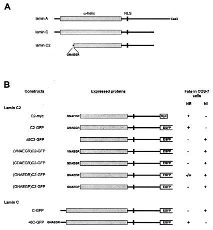
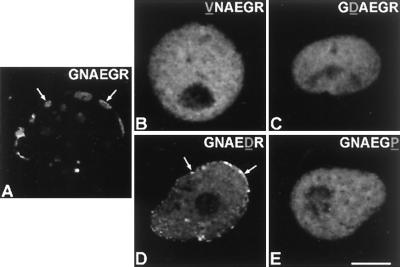
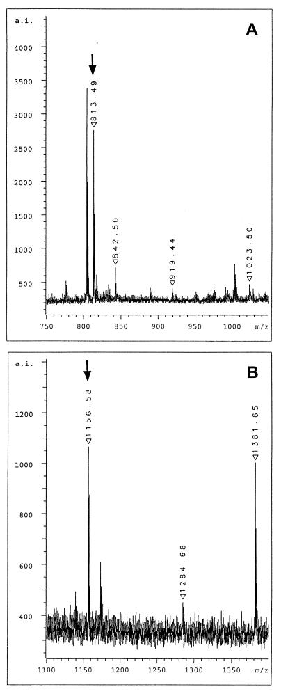
Meiotic lamin C2 is the only A-type lamin expressed during mammalian spermatogenesis. Typical for this short lamin is the unique hexapeptide GNAEGR, which substitutes the nonhelical amino terminus and part of the alpha-helical rod domain present in somatic lamins. Meiotic lamin C2 also lacks a carboxyl-terminal CaaX box, which is modified by isoprenylation and involved in nuclear envelope (NE) association of somatic isoforms. The mechanism by which lamin C2 becomes localized in the NE is totally unknown. Here we demonstrate that the hexapeptide GNAEGR is essential for this process: (i) Its deletion resulted in a diffuse distribution of lamin C2 within nuclei of transfected COS-7 cells; (ii) Mutated somatic lamin C, containing the sequence GNAEGR at its amino terminus, was located at the NE. The mass spectrometric analysis of the amino terminus of lamin C2 revealed that it is modified by myristoylation. Correspondingly, the substitution of the first glycine residue abolishes the NE association of lamin C2. We conclude that NE association of lamin C2 is achieved by a mechanism different from that of somatic lamins.


Figures

Figure 1
Figure 1
(A) Schematic representation of A-type lamins A, C, and C2. (B) Schematic representation and fate of the fusion proteins expressed in COS-7 cells. NI, nuclear interior.
Figure 2
Figure 2
Expression of somatic lamin C in cultured COS-7 cells as investigated by confocal laser scanning microscopy. (A and B) Protein C-GFP is localized within nuclei. (A′) The distribution in the same cell as in A of the transfected and endogenous lamins (L) is shown with the aid of mAb L3f4, which is specific for lamins A/C, B1, and B2. (B′) The distribution of DNA in the transfected cell shown in B is revealed by anti-DNA antibodies. Overlays are seen in (A" and B"). (Bar = 10 μm.)
Figure 3
Figure 3
Expression of meiotic lamin C2 in cultured COS-7 cells as investigated by confocal laser scanning microscopy. (A and B) In these cells, most lamin C2 (construct C2-GFP) is located at the nuclear periphery where it forms plaques. The expression level appears to be higher in the cell shown in B. (C) The fate of myc-tagged lamin C2 (construct C2-myc) is indistinguishable from that of lamin C2-GFP. The distribution of DNA in the same cells is revealed with the aid of an anti-DNA antibody in A′C′. Overlays are seen in A"C". (Bar = 10 μm.)
Figure 4
Figure 4
Role of hexapeptide GNAEGR in NE association as shown by confocal laser scanning microscopy. (A and B) Intranuclear distribution of mutant lamin C2 lacking the amino-terminal hexapeptide (construct Δ6C2-GFP). (C and D) Mutant lamin C carrying hexapeptide GNAEGR (construct + 6C-GFP) is located at the nuclear periphery. (A′) Endogenous lamins (L) are evidenced by using the mAb L3f4, an antibody that does not recognize lamin C2. (B′ and D′) The distribution of DNA is shown after incubation of the cells with a human anti-DNA antibody. (C′) The endogenous nuclear lamina structure is shown with the aid of the lamin B2-specific mAb X223. Overlays are seen in A"D". (Bar = 10 μm.)
Figure 5
Figure 5
Fate of lamin C2 as well as of lamin C2 carrying a mutated amino-terminal hexapeptide as investigated by confocal laser scanning microscopy. (A) Lamin C2 (construct C2-GFP) carrying the wild-type amino-terminal hexapeptide. Mutant lamins (VNAEGR)C2-GFP (B), (GDAEGR)C2-GFP (C), (GNAEDR)C2-GFP (D), and (GNAEGP)C2-GFP (E) are shown. Arrows denote some of the accumulations of expressed lamins that are located at the nuclear preriphery. (Bar = 10 μm.)
Figure 6
Figure 6
MALDI mass spectrometric analysis of total enzymatic digests of lamin C2. (A) Selected view of the mass spectrum of the tryptic digest. The ion signal at m/z 813.49 represents the myristoylated amino-terminal hexapeptide. (B) Selected view of the mass spectrum of the Lys-C digest. The ion signal at m/z 1156.58 represents the myristoylated amino-terminal peptide myrGNAEGRNTK. Arrows denote the ion signal of the myristoylated peptides.

References

    1. Burke B, Gerace L. Cell. 1986;44:639–652. - PubMed
    1. Benavente R, Krohne G. J Cell Biol. 1986;103:1847–1854. - PMC - PubMed
    1. Newport J W, Wilson K L, Dunphy W G. J Cell Biol. 1990;111:2247–2259. - PMC - PubMed
    1. Lenz-Böhme B, Wisnar J, Fuchs S, Reifegerste R, Buchner E, Betz H, Schmitt B. J Cell Biol. 1997;137:1001–1016. - PMC - PubMed
    1. Spann T P, Moir R D, Goldman A E, Stick R, Goldman R D. J Cell Biol. 1997;136:1201–1212. - PMC - PubMed

Publication types

LinkOut - more resources