. 2000 Nov 15;19(22):6020-9.
doi: 10.1093/emboj/19.22.6020.

Vezatin, a novel transmembrane protein, bridges myosin VIIA to the cadherin-catenins complex

Affiliations

Vezatin, a novel transmembrane protein, bridges myosin VIIA to the cadherin-catenins complex

P Küssel-Andermann et al. EMBO J. .

Abstract

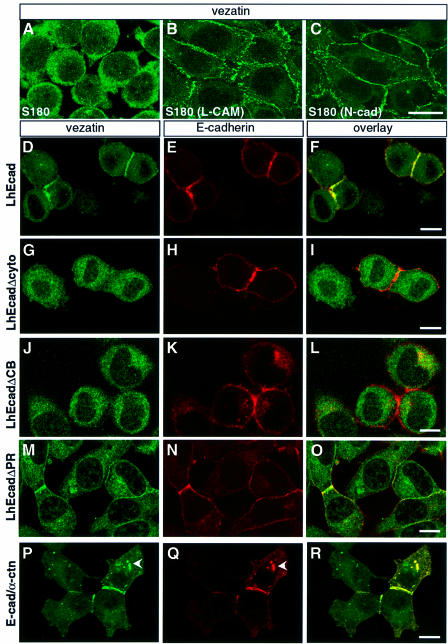
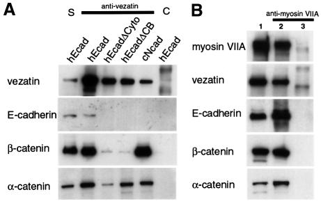
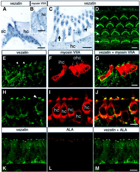
Defects in myosin VIIA are responsible for deafness in the human and mouse. The role of this unconventional myosin in the sensory hair cells of the inner ear is not yet understood. Here we show that the C-terminal FERM domain of myosin VIIA binds to a novel transmembrane protein, vezatin, which we identified by a yeast two-hybrid screen. Vezatin is a ubiquitous protein of adherens cell-cell junctions, where it interacts with both myosin VIIA and the cadherin-catenins complex. Its recruitment to adherens junctions implicates the C-terminal region of alpha-catenin. Taken together, these data suggest that myosin VIIA, anchored by vezatin to the cadherin-catenins complex, creates a tension force between adherens junctions and the actin cytoskeleton that is expected to strengthen cell-cell adhesion. In the inner ear sensory hair cells vezatin is, in addition, concentrated at another membrane-membrane interaction site, namely at the fibrillar links interconnecting the bases of adjacent stereocilia. In myosin VIIA-defective mutants, inactivity of the vezatin-myosin VIIA complex at both sites could account for splaying out of the hair cell stereocilia.


Figures

None
Fig. 1. Schematic representation of vezatin splice variants. For each cDNA the open reading frame (ORF) is represented by a grey box and the putative transmembrane domain (TM), when present, by a black box. The A34 fragment (dashed line) that was identified by the yeast two-hybrid screen is indicated (A34 prey). Two cDNA clones containing complete ORFs, A34.1 and A34.2, were obtained. The A34.1 sequence predicts an ATG initiator codon preceded by a stop codon 18 nt upstream. It encodes a putative 569 amino acid protein, with a transmembrane domain (amino acids 96–115) but no signal peptide. A34.1 is used as a reference for nucleotide and amino acid positions. A34.2 has a distinct predicted ATG initiator codon preceded by a stop codon 6 nt upstream. The deduced 460 amino acid protein sequence lacks the TM segment; it differs from the A34.1 sequence by the N-terminal 10 amino acids. These two cDNAs were also present in a human retinal library. In addition, partial cDNA sequences were obtained. A34.3 and A34.5 cDNAs, which were obtained by 5′-RACE, contain short in-frame deletions (339 nt in A34.3 and two deletions of 57 and 73 nt in A34.5) in the extracellular region. A34.4, A34.5 and EST AA432068 carry a 73 nt deletion (nt 134–206) encompassing the ATG initiator codon of A34.1, thus resulting in an upstream extension of the ORF. In A34.2 and A34.3 cDNAs a 12 nt in-frame insertion (+12) at nt position 749 (amino acid 249) was observed. In A34.6 and several ESTs (see Materials and methods) a 1072 nt deletion (–1072), covering nt 1869–2940 of A34.1, results in an ORF extension of 182 amino acids into the 3′-untranslated region. Finally, EST AA432068 encodes a putative vezatin variant with a short cytoplasmic tail, which lacks the myosin VIIA interacting domain (nt 1188–1889). Stars indicate the positions of the two human peptides, A34P1 and A34P2, used to generate antibodies.
None
Fig. 2. Vezatin binds to the C-terminal FERM domain of myosin VIIA. (A) Expression of vezatin in adult mouse tissues. Total protein extracts (10 µg/lane) from six different tissues of 10-day-old mice were immunoblotted with the anti-mA34 antibody. Multiple vezatin isoforms are observed in all tissues. The higher bands may correspond to vezatin isoforms with large extracellular domains (see Figure 1). (B) Binding of vezatin to the myosin VIIA tail in co-transfected HEK293 cells. Extracts from co-transfected cells expressing both the myc-tagged A34.2 peptide and the myosin VIIA tail (lane 3) were used for co-immunoprecipitation experiments; the myosin VIIA tail and A34.2 peptide are co-immunoprecipitated with either the anti-myosin VIIA (lane 4) or the anti-myc (lane 5) antibody. When using extracts from HEK293 cells producing the myc-tagged A34.2 peptide alone (lane 1), no immunoprecipitate forms with the anti-myosin VIIA antibody (lane 2). (C) Binding of vezatin to the myosin VIIA C-terminal FERM domain. A standard amount of Caco-2 cell lysate containing endogenous vezatin (5% of which is shown in lane SN) was incubated with avidin resins coated with different biotinylated myosin VIIA peptides (bait, amino acids 1752–2215; BdC2, 1752–1931; BdN1, 1896–2215) or a biotinylated control protein, chloramphenicol acetyltransferase (CAT). Vezatin binds to either the bait peptide or the BdN1 fragment containing only the FERM domain, but not to BdC2 lacking the FERM domain or to CAT.
None
Fig. 3. Co-localization of endogenous vezatin and the myosin VIIA tail fused to GFP in transfected MDCK cells. As soon as cell contacts can be detected, the myosin VIIA tail (A) co-localizes with endogenous vezatin (B) at the precise membrane sites of the cell–cell contacts (C). The same result was obtained with a GFP–myosin VIIA tail fragment composed of the last 464 amino acids of myosin VIIA (not shown). After Triton X-100 treatment the GFP–myosin VIIA tail (D) is still associated with the actin complex (E) at the cell–cell junctions (F). Bar, 10 µm.
None
Fig. 4. Vezatin at cell–cell contacts. (AC) Expression of vezatin in adult mouse tissues. (A) Oblique section of a P20 mouse intestinal villosity. The vezatin immunoreactivity is mainly localized at the cell–cell junctions of the epithelial cells. The connective tissue is also immunoreactive. (B) P20 mouse skin. The bulk of the vezatin immunoreactivity is located between epidermal cells. A punctate labelling is also detected in the derma. (C) P20 mouse lung. An intense vezatin staining is observed between the epithelial cells of the bronchial tube. (DJ) Co-localization of vezatin with E-cadherin at cell–cell contacts. MDCK cells were grown to either semi-confluence (D–F) or confluence (G–J). Vezatin (D and G) and E-cadherin (E and H) co-localize at cell–cell contacts (F, I and J). (J) A computer-generated Z view (vertical axis) reconstructed from a series of cross-sections, showing the co-distribution of vezatin and E-cadherin along the lateral cell membrane. Co-localizations in the double staining experiments are shown on merged images (F, I and J). Bar, 10 µm.
None
Fig. 5. Vezatin interacts with the cadherin–catenin complex. (AC) Localization of vezatin in S180 cells expressing chicken E-cadherin (L-CAM) or N-cadherin (N-cad). In S180 fibroblasts (A), which lack E-cadherin and adherens-like junctions, vezatin is distributed throughout the cytoplasm. In contrast, in stably transfected S180 cells expressing chicken L-CAM (B) or N-cadherin (C) an intense vezatin labelling is detected at cell–cell contacts. (DR) Vezatin localization in transfected L2071 cells expressing different truncated E-cadherin (D–O) or E-cadherin–α-catenin chimeras (P–R). In L cells stably transfected with the entire human E-cadherin (L-hEcad), vezatin (D) as well as E-cadherin (E) is recruited to the cell–cell contacts (F). In contrast, in L cells stably expressing hEcad lacking either the cytoplasmic domain (L-hEcadΔCyto) (G–I) or the β-catenin binding domain (L-hEcadΔCB) (J–L) vezatin has a cytoplasmic localization. In L cells expressing hEcad lacking the p120-catenin binding site (L-hEcadΔPR) vezatin is recruited to the cell junction (M–O). In L cells expressing the E-cadherin–α-catenin chimera, linking the E-cadherin transmembrane domain directly to the last 398 C-terminal amino acids of α-catenin, vezatin is detected at the cell–cell junctions (P–R). Note the cytoplasmic localizations of vezatin and E-cadherin–α-catenin chimera (arrowheads). Bar, 10 µm.
None
Fig. 6. Immunoprecipitation experiments in transfected cells expressing different forms of E-cadherin. (A) Extracts from L cells expressing the entire human E-cadherin (hEcad), human E-cadherin lacking the cytodomain (hEcadΔCyto) or the β-catenin binding domain (hEcadΔCB) and from S180 cells expressing the entire chicken N-cadherin (cNcad) were used. Vezatin, α-catenin and β-catenin are co-immunoprecipitated by an anti-vezatin antibody in cells transfected with either E- or N-cadherin cDNAs. In addition, vezatin and α-catenin are co-immunoprecipitated in cells producing the truncated E-cadherin variants (lanes hEcadΔCyto and hEcadΔCB), whereas only a very small fraction of β-catenin, compared with control (hEcad), is detected in the immunoprecipitate. The preimmune serum (lane C) was used as a negative control. S, soluble fraction. (B) In extracts from HEK293 cells expressing myosin VIIA tail (lane 2) the myosin VIIA tail co-immunoprecipitates with vezatin, E-cadherin, β-catenin and α-catenin, using an anti-myosin VIIA antibody. No immuno precipitation was observed with extracts from non-transfected cells (lane 3). Lane 1 contains the soluble fraction.
None
Fig. 7. Co-localization of vezatin, myosin VIIA and the ankle link antigen (ALA) in the inner ear. (AC) Immunoelectron microscopy of the mouse cochlea, showing the ultrastructural distribution of vezatin and myosin VIIA. (A and B) Oblique sections through the junction between a hair cell (hc) and a supporting cell (sc) at P30, showing vezatin (A) and myosin VIIA (B) labelling along the adherens junction. (C) In the hair bundle vezatin labelling is concentrated at the base (arrowhead) of the stereocilia, where the ankle links (arrow) are located. (DM) Immunohistofluorescence. (D) Whole mount preparation of a P2 organ of Corti. Vezatin immunoreactivity is observed in the stereocilia hair bundles of the three rows (arrowheads) of outer hair cells (W shape) and the single row (arrow) of inner hair cells (U shape). (E–G) P2 mouse cochlea (fixed with PFA). (E) Vezatin is detected in the inner (ihc) and outer (ohc) hair cells of the organ of Corti. Note that the hair bundles are strongly immunoreactive (arrowheads). Vezatin is also detected in the supporting cells and nerve fibres. When either unfixed or methanol fixed sections were used a strong vezatin labelling was observed also at the cell–cell contacts between hair cells and supporting cells (not shown). (F) Myosin VIIA is restricted to the ihc and ohc, where it is localized throughout the cell. (H–J) P8 mouse utricular hair cells (hc). (H) The strongest vezatin immunoreactivity is at the base of the stereocilia (arrowhead), whereas the myosin VIIA labelling (I) appears more uniform. (K–M) Adult chicken inner ear macula. Vezatin (K) and the ankle link antigen ALA (L) are co-localized (M) at the base of the stereocilia. Bars: (A) and (B) 0.1 µm; (C) 0.5 µm; (D)–(M) 10 µm.

References

    1. Aberle H., Butz,S., Stappert,J., Weissig,H., Kemler,R. and Hoschuetzky,H. (1994) Assembly of the cadherin-catenin complex in vitro with recombinant proteins. J. Cell Sci., 107, 3655–3663. - PubMed
    1. Adams C.L. and Nelson,W.J. (1998) Cytomechanics of cadherin-mediated cell–cell adhesion. Curr. Opin. Cell Biol., 10, 572–577. - PubMed
    1. Adams C.L., Nelson,W.J. and Smith,S.J. (1996) Quantitative analysis of cadherin-catenin–actin reorganization during development of cell–cell adhesion. J. Cell Biol., 135, 1899–1911. - PMC - PubMed
    1. Adams C.L., Chen,Y.T., Smith,S.J. and Nelson,W.J. (1998) Mechan isms of epithelial cell–cell adhesion and cell compaction revealed by high-resolution tracking of E-cadherin–green fluorescent protein. J. Cell Biol., 142, 1105–1119. - PMC - PubMed
    1. Angres B., Barth,A. and Nelson,W.J. (1996) Mechanism for transition from initial to stable cell–cell adhesion: kinetic analysis of E-cadherin-mediated adhesion using a quantitative adhesion assay. J. Cell Biol., 134, 549–557. - PMC - PubMed

Publication types

MeSH terms