. 2000 Nov 15;19(22):6075-84.
doi: 10.1093/emboj/19.22.6075.

Phosphorylation and N-terminal region of yeast ribosomal protein P1 mediate its degradation, which is prevented by protein P2

Affiliations

Phosphorylation and N-terminal region of yeast ribosomal protein P1 mediate its degradation, which is prevented by protein P2

G Nusspaumer et al. EMBO J. .

Abstract

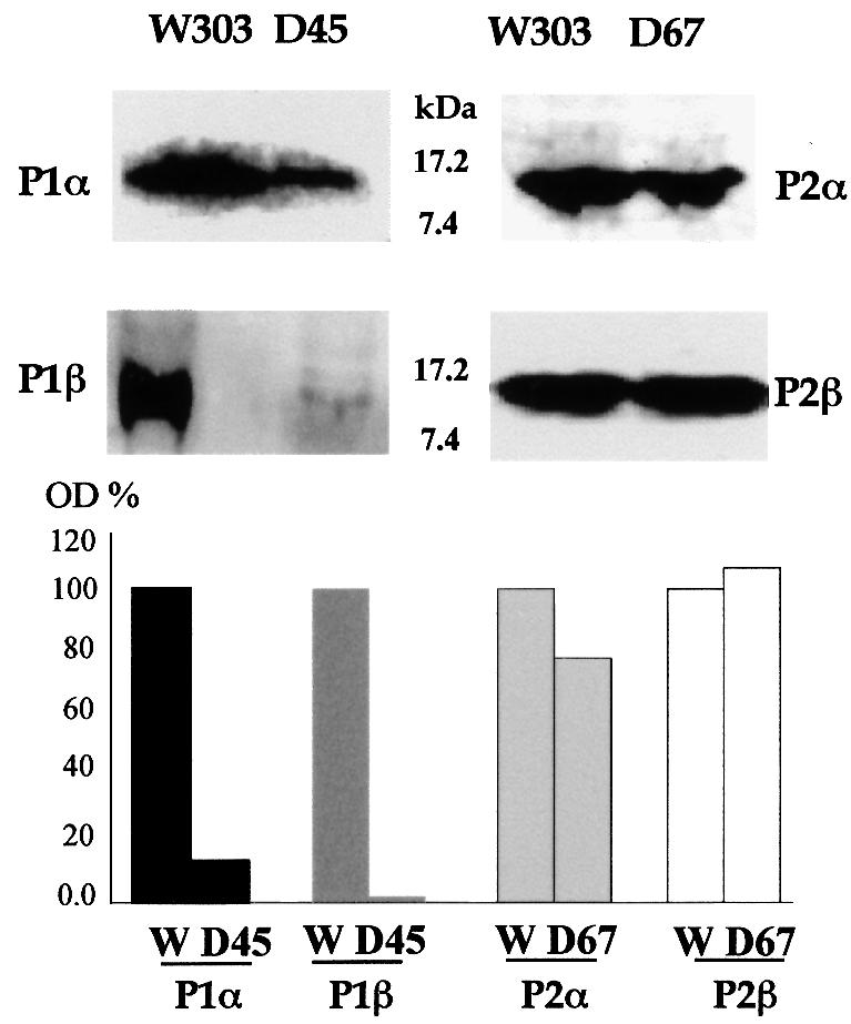
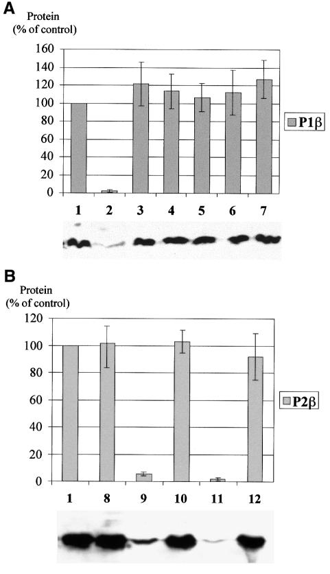
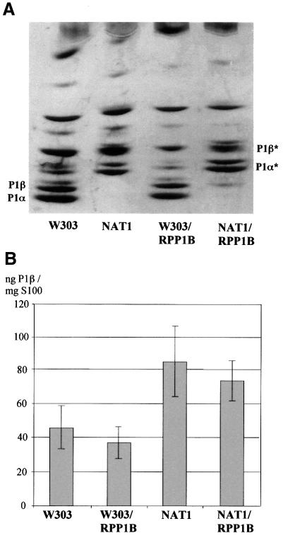
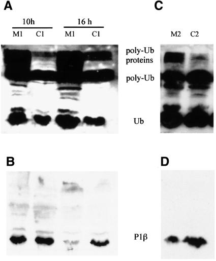
The stalk proteins P1 and P2, which are fundamental for ribosome activity, are the only ribosomal components for which there is a cytoplasmic pool. Accumulation of these two proteins is differentially regulated in Saccharomyces cerevisiae by degradation. In the absence of P2, the amount of P1 is drastically reduced; in contrast, P2 proteins are not affected by a deficiency in P1. However, association with P2 protects P1 proteins. The half-life of P1 is a few minutes, while that of P2 is several hours. The proteasome is not involved in the degradation of P1 proteins. The different sensitivity to degradation of these two proteins is associated with two structural features: phosphorylation and N-terminus structure. A phosphorylation site at the C-terminus is required for P1 proteolysis. P2 proteins, despite being phosphorylated, are protected by their N-terminal peptide. An exchange of the first five amino acids between the two types of protein makes P1 resistant and P2 sensitive to degradation.


Figures

None
Fig. 1. Immunoblot analysis of P1/P2 levels in S.cerevisiae W303-1b, D45 and D67 strains. Total extracts (100 µg) from cells grown to mid-logarithmic phase in rich medium were separated by 15% SDS–PAGE followed by immunoblot analysis using monoclonal antibodies to P1β (1CE1), P2α (1BE3) and P2β (1AA9) and a rabbit polyclonal antibody to P1α. Densitometric estimation of the intensity of the bands was performed; results are shown as a percentage of the S.cerevisiae W303-1b band intensity.
None
Fig. 2. Estimation of P1β mRNA in S.cerevisiae W303-1b and D45 by a northern assay. Total RNA was resolved by electrophoresis, blotted onto a membrane and detected by hybridization using a 32P-labelled 1.3 kb PstI–HindIII DNA fragment containing the RPP1B coding sequence derived from pMRH46 (Remacha et al., 1988). A 1.0 kb 32P-labelled AvaI–HindIII DNA fragment from pYactII containing actin gene ACT1 was used as a standard. The results of the densitometric estimation are plotted as an RPP1B/ACT1 ratio.
None
Fig. 3. Half-life of P1β and P2β proteins estimated by pulse–chase labelling. Saccharomyces cerevisiae D45/RPP1B and D67 were labelled with [35S]Met–Cys for 10 min and then chased with an excess of cold methionine and cysteine. At the time points indicated, cell aliquots were withdrawn, and the amount of P1β (closed circles) and P2β (open circles) proteins was estimated in D45/RPP1B and D67, respectively, by immunoprecipitation and SDS–PAGE as indicated in Materials and methods.
None
Fig. 4. Stability of acidic proteins determined by cycloheximide inhibition. Saccharomyces cerevisiae strains D45/RPP1B, D45/RPP1A and D67 growing exponentially in SC medium were treated with cycloheximide to inhibit protein synthesis. Aliquots were taken at the indicated times after the addition of cycloheximide and the amount of proteins in the cell extracts was estimated by western blotting using the corresponding specific antibodies (see Figure 1).
None
Fig. 5. Effect of N-terminus and phosphorylation on accumulation of acidic proteins. (A) Effects on P1β. P1β chimeric genes carrying different modifications as listed below, cloned in pFL36, were expressed in S.cerevisiae D456. Strains W303-1b (1) and D456/111 (2) were included as controls. The amount of P1β protein in the extracts was estimated by western blotting using monoclonal antibody 1CE1. P1β modifications: (3) P1β(10N2β), first 10 amino acids from P2β; (4) P1β(5N2β), first five amino acids from P2β; (5) P1β(S96C), Ser96 mutated to cysteine; (6) P1β(S96F), Ser96 mutated to phenylalanine; (7) P1β(5N2β/S96C), first five amino acids from P2β and Ser96 mutated to cysteine. A representative western blot experiment is shown below the histogram. Error bars show the standard deviation of three independent experiments. (B) Effects on P2β. Modified P2β genes cloned in pFL38 were expressed in S.cerevisiae D567. Strains W303-1b (1) and D567/222 (8) were used as controls. Samples were processed as in (A) and protein P2β was assayed using monoclonal antibody 1AA9. P2β modifications: (9) P2β(10N1β), first 10 amino acids from P1β; (10) P2β(10N1β/S100F), first 10 amino acids from P1β and Ser100 mutated to phenylalanine; (11) P2β(5N1β), first five amino acids from P1β; (12) P2β(5N1β/S100F), first five amino acids from P1β and Ser100 mutated to phenylalanine. A representative western blot experiment is shown below the histogram. Error bars show the standard deviation of three independent experiments.
None
Fig. 6. Effect of N-acetyltransferase (NAT1) inactivation on P1β protein accumulation. (A) Ribosomes from untransformed S.cerevisiae W303-1b and NAT1, and from the same two strains transformed with plasmid YEp356/RPP1B, were resolved by isoelectrofocusing in the pH range 2.0–5.0. Proteins were silver stained. P1α* and P1β* mark the positions of the non-acetylated forms. (B) The amount of free P1β protein in these strains was estimated by inhibition ELISA in cell extracts deprived of ribosomes (S100 fractions). Error bars show the standard deviation of three independent experiments.
None
Fig. 7. P1β degradation is proteasome independent. Gene dosage experiments were carried out by overexpressing P1β under restrictive conditions in proteasomal mutant strains FS11-1b (M1) and WCG4a-11/21 (M2), and in parental strains FY1679 (C1) and WCG4a (C2). In the case of FS11-1b, S100 extracts were prepared 10 or 16 h after the change of medium (A and B). For WCG4a-11/21, S100 fractions were prepared after 5 h at 38°C in mineral medium (C and D). Western blot analysis using anti-ubiquitin (A and C) and anti-P1β (B and D) antibodies was performed.

References

    1. Ballesta J.P.G. and Remacha,M. (1996) The large ribosomal subunit stalk as a regulatory element of the eukaryotic translational machinery. Prog. Nucleic Acid Res. Mol. Biol., 55, 157–193. - PubMed
    1. Ballesta J.P.G., Guarinos,E., Zurdo,J., Parada,P., Nusspaumer,G., Lalioti,V.S., Perez-Fernandez,J. and Remacha,M. (2000) Structure of the yeast ribosomal stalk. In Garrett,R., Douthwaite,S., Matheson,A., Liljas,A., Moore,P.B. and Noller,H.F. (eds), The Ribosome. Structure, Function, Antibiotics and Cellular Interactions. American Society for Microbiology, Washington, DC, pp. 115–125.
    1. Bonneaud N., Ozier-Kalogeropoulos,O., Li,G.Y., Labouesse,M., Minvielle-Sebastia,L. and Lacroute,F. (1991) A family of low and high copy replicative, integrative and single-stranded S. cerevisiae/E. coli shuttle vectors. Yeast, 7, 609–615. - PubMed
    1. Broach J., Strathern,J.N. and Hicks,J.B. (1979) Transformation in yeast: development of a hybrid cloning vector and isolation of the CAN1 gene. Gene, 8, 121–133. - PubMed
    1. Bromley S., Hereford,L. and Rosbash,M. (1982) Further evidence that the rna2 mutation of Saccharomyces cerevisiae affects mRNA processing. Mol. Cell. Biol., 2, 1205–1211. - PMC - PubMed

Publication types

MeSH terms