. 2000 Nov 15;19(22):6085-97.
doi: 10.1093/emboj/19.22.6085.

Nuclear-specific degradation of Far1 is controlled by the localization of the F-box protein Cdc4

Affiliations

Nuclear-specific degradation of Far1 is controlled by the localization of the F-box protein Cdc4

M Blondel et al. EMBO J. .

Abstract

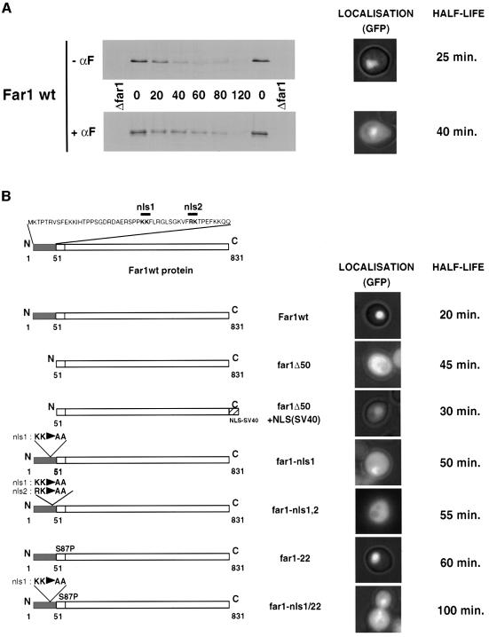
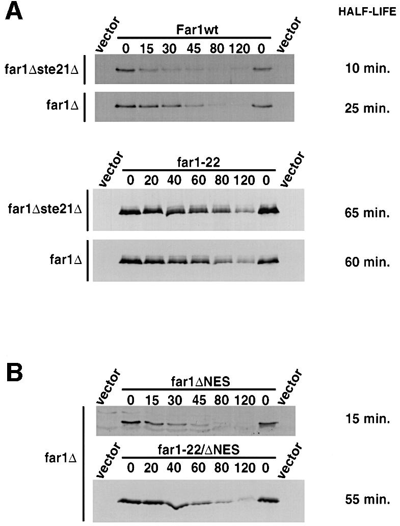
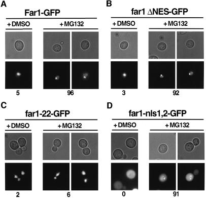
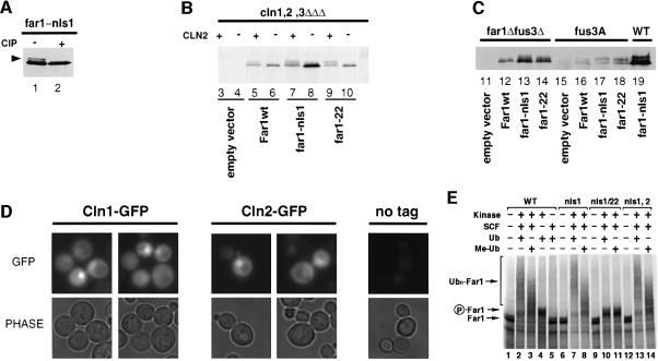
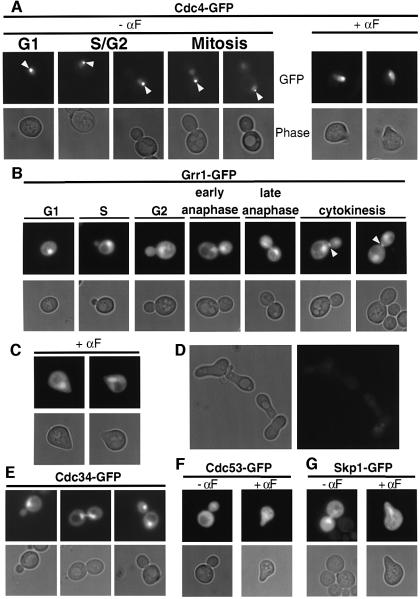
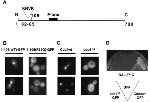
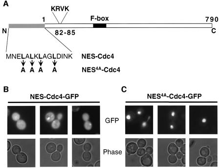
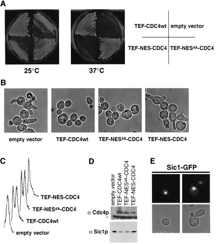
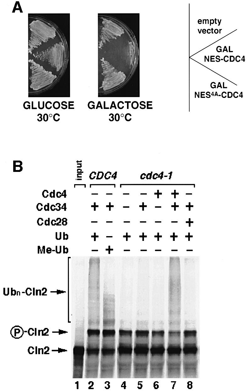
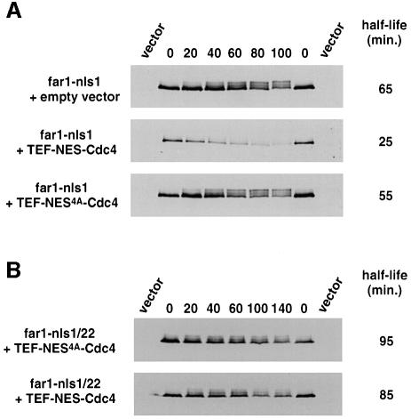
Far1 is a bifunctional protein that is required to arrest the cell cycle and establish cell polarity during yeast mating. Here we show that SCF(Cdc4) ubiquitylates Far1 in the nucleus, which in turn targets the multi-ubiquitylated protein to 26S proteasomes most likely located at the nuclear envelope. In response to mating pheromones, a fraction of Far1 was stabilized after its export into the cytoplasm by Ste21/Msn5. Preventing nuclear export destabilized Far1, while conversely cytoplasmic Far1 was stabilized, although the protein was efficiently phosphorylated in a Cdc28-Cln-dependent manner. The core SCF subunits Cdc53, Hrt1 and Skp1 were distributed in the nucleus and the cytoplasm, whereas the F-box protein Cdc4 was exclusively nuclear. A cytoplasmic form of Cdc4 was unable to complement the growth defect of cdc4-1 cells, but it was sufficient to degrade Far1 in the cytoplasm. Our results illustrate the importance of subcellular localization of F-box proteins, and provide an example of how an extracellular signal regulates protein stability at the level of substrate localization.


Figures

None
Fig. 1. Cytoplasmic Far1 is stabilized. (A) The half life of Far1 expressed from the GAL promoter was determined by GAL shut-off experiments (see Material and methods) in far1Δ cells (YMP1054) treated (lower blot) or not treated (upper blot) with α-factor (αF) for 2 h. The blots were quantified and the half life (in min) is indicated on the right. The right panels show the localization of Far1–GFP under the same conditions. (B) The half lives (in min) of wild-type and Far1 mutants schematically indicated on the left were determined by GAL shut-off experiments. The localization of the corresponding GFP fusion proteins was visualized by fluorescence microscopy and was overlaid with the phase contrast image.
None
Fig. 2. Preventing nuclear export of Far1 increases its rate of degradation. (A) The half lives of wild-type Far1 (upper panels) or Far1-22 (lower panels) were determined in far1Δste21Δ (YMP1067, upper blots) or for control Δfar1 cells (YMP1054, lower blots) by the GAL shut-off protocol as described. (B) The half lives of Far1-ΔNES (far1ΔNES, upper panel) or Far1-22/ΔNES (far1-22/ΔNES, lower panel) were determined by GAL shut-off experiments in far1Δ cells.
None
Fig. 3. Ubiquitylated Far1 accumulates at specific sites in the nucleus. (ADerg6Δ cells (RH3622) expressing the indicated wild-type or mutant Far1 fused to GFP were treated with the proteasome inhibitor MG132 (+ MG132) or, for control, its solvent DMSO (+ DMSO) and visualized after 3 h by phase contrast (upper rows) or fluorescence microscopy (lower rows). The numbers indicate the percentage of cells that accumulated GFP spots; at least 200 cells were counted for each strain.
None
Fig. 4. Cdc28–Cln-dependent phosphorylation of Far1 can occur in the cytoplasm and the nucleus. (A) Far1-nls1 accumulates phosphorylated species (arrowhead). Extracts from far1Δ cells expressing Far1-nls1 were incubated with active (+ CIP, lane 2) or heat-inactivated (– CIP, lane 1) alkaline phosphatase, and immunoblotted with specific antibodies. (B) YMG258 cells (cln1,2,3Δ MET-CLN2) transformed with an empty control plasmid (lanes 3 and 4), or plasmids expressing wild-type (lanes 5 and 6) or mutant Far1 (lanes 7–10) as indicated, were grown in selective media lacking methionine at 30°C to early log phase; the culture was divided and methionine was added to one half to repress Cln2 expression (– Cln2), while no methionine was added to the control half (+ Cln2). Extracts were prepared after 3 h and immunoblotted for Far1. (C) The phosphorylation of wild-type or mutant Far1 was analyzed as above in either far1Δ (YMP1054; lane 19), far1Δ fus3Δ (YMP291, lanes 11–14) or fus3A cells (YMP293, lanes 16–18), transformed with an empty control vector (lanes 11 and 15) or plasmids expressing either wild-type (lanes 12 and 16) or the indicated mutant Far1 (lanes 13, 14, 17–19). (D) Cln1–GFP and Cln2–GFP are localized both in the cytoplasm and in the nucleus. Wild-type cells expressing either Cln1–GFP (left panels) or Cln2–GFP (middle panels) from the GAL promoter were grown at 30°C to early log phase in selective media, and analyzed by fluorescence microscopy. The right panel (no GFP) shows cells expressing untagged Cln2 to control for background fluorescence. (E) Ubiquitylation of wild-type and mutant Far1 by SCFCdc4 in vitro. Wild type (lanes 1–5) or the indicated Far1 mutants (lanes 6–14) were in vitro translated in the presence of [35S]methionine and incubated as indicated with reconstituted SCFCdc4 (SCF), Cdc28–Cln2 (Kinase) and ubiquitin (Ub) as described in Materials and methods. Where indicated, Me-ubiquitin instead of ubiquitin was added as a control (Me-Ub). The arrows point to the position of unphosphorylated (Far1) and phosphorylated Far1 (P-Far1), respectively, while the brackets mark the position of multi-ubiquitylated Far1 (Ubn-Far1).
None
Fig. 5. Cdc4–GFP is exclusively in the nucleus while Grr1–GFP is localized in both nucleus and cytoplasm. (A) Wild-type cells (K699) expressing Cdc4–GFP from the GAL promoter were grown to early log phase in selective media at 30°C, and analyzed by phase (lower rows) and fluorescence microscopy (upper rows). Where indicated, cells were treated with pheromones for 2 h (+ α-factor, right panels). Note that Cdc4–GFP is nuclear at all stages of the cell cycle and in response to pheromones, and may localize to the mother-specific spindle pole body (arrowhead). (BDgrr1Δ cells (YMP2957) transformed with a control plasmid (D) or a plasmid expressing Grr1–GFP from the GAL promoter were analyzed as described in (A). Grr1–GFP is found at the cytokinesis ring late in mitosis (B, arrowhead). (EG) Wild-type cells (K699) transformed with plasmids expressing either Cdc34–GFP (E), Cdc53–GFP (F) or Skp1–GFP (G) from the GAL promoter were analyzed as described in (A). Where indicated, α-factor was added for 2 h.
None
Fig. 6. Cdc4 contains a monopartite NLS in its N-terminus. (A) Schematic representation of Cdc4. The monopartite NLS (amino acids 82–85; KRVK) and the F-box (black bar) are indicated. The numbers indicate the amino acids starting from the N-terminus (N) to the C-terminus (C) of Cdc4. (B) Wild-type cells (K699) expressing from the GAL promoter GFP fused to the N-terminal 106 amino acids of wild-type (Cdc41–106–GFP, left panels) or mutant Cdc4 (Cdc41–106(R83G)–GFP, right panels) were analyzed by fluorescence microscopy. (C) Wild-type cells (K699) expressing from the GAL promoter either wild-type Cdc4–GFP (Cdc4wt, left panels) or a mutant Cdc4–GFP harboring three changes (K82A, R83A and K85A) in the putative monopartite NLS (Cdc43A, right panels). (Dcdc4-1 cells expressing GFP alone (GFP), Cdc43A–GFP or wild-type Cdc4–GFP from the GAL promoter were grown for 3 days at 37°C on media containing galactose (GAL).
None
Fig. 7. A fusion of Cdc4 with the NES of human PKI is predominantly cytoplasmic. (A) Amino acid sequence of the wild-type (NES–Cdc4) or mutant NES sequence (NES4A–Cdc4) fused to the N-terminus of Cdc4. (B and C) GFP fluorescence (upper rows) and phase microscopy images (lower rows) of wild-type cells (K699) expressing NES–Cdc4–GFP (B) or NES4A–Cdc4–GFP (C) from the inducible GAL promoter. Cells were grown at 30°C to early log phase in selective media containing 2% raffinose, at which time expression of NES–Cdc4–GFP or NES4A–Cdc4–GFP was induced for 3 h by addition of 2% galactose.
None
Fig. 8. Expression of NES–Cdc4 is not able to bypass the G1 arrest of cdc4-1 mutant cells. (Acdc4-1 cells (YMT668) were transformed with an empty control vector or plasmids expressing from the constitutive TEF promoter either NES4A–Cdc4, NES–Cdc4 or wild-type Cdc4, and grown for 3 days on selective media at either 25°C (left panel) or 37°C (right panel). (B and C) To examine the arrest phenotype, the cdc4-1 transformants described above were grown at 25°C to early log phase in selective media, at which time the cultures were shifted to 37°C. After 3 h, the cells were analyzed by phase contrast microscopy (B) or FACS analysis (C). (D) An aliquot of the cultures described in (B) was analyzed by immunoblotting for the presence of Cdc4 (upper blot) and Sic1 (lower blot). The asterisk marks the position of a protein that cross reacts with the Cdc4 antibody. (E) GFP fluorescence (upper rows) and phase microscopy images (lower rows) of wild-type cells (K699) expressing Sic1–GFP.
None
Fig. 9. Subcellular localization may contribute to substrate specificity of Cdc4 in vivo. (A) Wild-type cells (K699) were transformed with a control vector (empty vector), or plasmids expressing either NES–Cdc4 or NES4A–Cdc4 from the inducible GAL promoter. Individual transformants were grown at 30°C on medium containing galactose (right plate, GAL promoter on) or glucose (left plate, GAL promoter off), and photographed after 3 days. (B) SCFCdc4 is able to ubiquitylate Cln2 in vitro. In vitro-translated [35S]methionine-labeled Cln2 (lane 1) was incubated in DEAE-extracts prepared from wild-type (lanes 2 and 3) or cdc4-1 cells (lanes 4–8), complemented where indicated (+) with ubiquitin (Ub), and bacculo-expressed Cdc34, Cdc4 and Cdc28. In lane 3, methylated ubiquitin (Me-Ub) was added instead of ubiquitin to compete for polyubiquitylation. The arrow points to unphosphorylated (Cln2) or phosphorylated Cln2 (P-Cln2), while the bracket marks the position of polyubiquitylated Cln2 (Ubn-Cln2).
None
Fig. 10. Wild-type cells expressing NES–Cdc4 are able to degrade cytoplasmic Far1-nls1. (A and B) The half life of Far1-nls1 (A) or Far1-22/nls1 (B) was determined by GAL-shut-off experiments in far1Δ cells (YMP1054) containing either an empty vector or plasmids expressing NES–Cdc4 or NES4A–Cdc4 from the TEF promoter. The blots were quantified and the half lives (in min) are shown on the right. Note that cytoplasmic NES–Cdc4 is able to degrade cytoplasmic Far1-nls1 in a manner dependent on a phosphorylatable serine 87.

References

    1. Ashcroft M., Taya,Y. and Vousden,K.H. (2000) Stress signals utilize multiple pathways to stabilize p53. Mol. Cell. Biol., 20, 3224–3233. - PMC - PubMed
    1. Bai C., Sen,P., Hofmann,K., Ma,L., Goebl,M., Harper,J.W. and Elledge,S.J. (1996) SKP1 connects cell cycle regulators to the ubiquitin proteolysis machinery through a novel motif, the F-box. Cell, 86, 263–274. - PubMed
    1. Barral Y., Jentsch,S. and Mann,C. (1995) G1 cyclin turnover and nutrient uptake are controlled by a common pathway in yeast. Genes Dev., 9, 399–409. - PubMed
    1. Baumeister W., Walz,J., Zuhl,F. and Seemuller,E. (1998) The proteasome: paradigm of a self-compartmentalizing protease. Cell, 92, 367–380. - PubMed
    1. Blondel M., Alepuz,P.M., Huang,L.S., Shaham,S., Ammerer,G. and Peter,M. (1999) Nuclear export of Far1p in response to pheromones requires the export receptor Msn5p/Ste21p. Genes Dev., 13, 2284–2300. - PMC - PubMed

Publication types

MeSH terms

Substances