. 2000 Nov 15;19(22):6121-30.
doi: 10.1093/emboj/19.22.6121.

Pontin52 and reptin52 function as antagonistic regulators of beta-catenin signalling activity

Affiliations

Pontin52 and reptin52 function as antagonistic regulators of beta-catenin signalling activity

A Bauer et al. EMBO J. .

Abstract

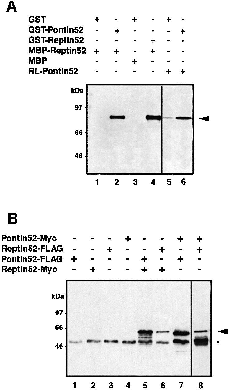
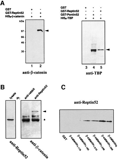
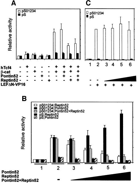
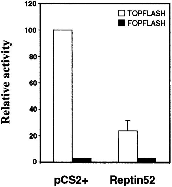
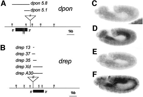
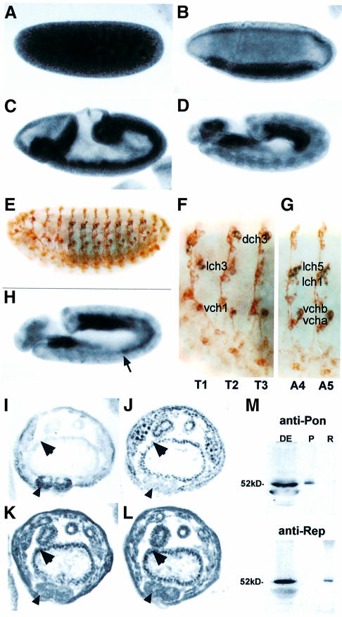
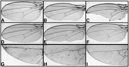
In Wnt-stimulated cells, beta-catenin becomes stabilized in the cytoplasm, enters the nucleus and interacts with HMG box transcription factors of the lymphoid-enhancing factor-1 (LEF-1)/T-cell factor (TCF) family, thereby stimulating the transcription of specific target genes. We recently identified Pontin52 as a nuclear protein interacting with beta-catenin and the TATA-box binding protein (TBP), suggesting its involvement in regulating beta-catenin-mediated transactivation. Here, we report the identification of Reptin52 as an interacting partner of Pontin52. Highly homologous to Pontin52, Reptin52 likewise binds beta-catenin and TBP. Using reporter gene assays, we show that the two proteins antagonistically influence the transactivation potential of the beta-catenin-TCF complex. Furthermore, we demonstrate the evolutionary conservation of this mechanism in Drosophila. dpontin and dreptin are essential genes that act antagonistically in the control of Wingless signalling in vivo. These results indicate that the opposite action of Pontin52 and Reptin52 on beta-catenin-mediated transactivation constitutes an additional mechanism for the control of the canonical Wingless/Wnt pathway.


Figures

None
Fig. 1. Homotypic and heterotypic interactions of Reptin52 and Pontin52 in vitro and in vivo. (AIn vitro association of Reptin52 with Pontin52 demonstrated by affinity precipitation of MBP–Reptin52 with GST–Pontin52 (lane 2). Self-association of Reptin52 and Pontin52 was detected analogously by precipitation with GST–Reptin (lane 4) and GST–Pontin (lane 6). Lanes 1–4, western blot revealed by anti-MBP antibodies; lanes 5 and 6, autoradiography of in vitro translated [35S]Pontin52 (RL-Pontin52). Arrow, precipitated Reptin52 or Pontin52. (BIn vivo association of Reptin52 with Pontin52 (lanes 5 and 8) and self-association of both proteins (lanes 6 and 7). Different combinations of Myc- and FLAG-tagged variants of Reptin52 and Pontin52 were transiently transfected in HEK293 cells. Western blot analysis of cell lysates immunoprecipitated with anti-Myc (lanes 1–7) and anti-FLAG antibodies (lane 8). Arrow, precipitated Myc-Pontin52 or Myc-Reptin52; asterisk, antibody heavy chain.
None
Fig. 2. Reptin52 interacts with β-catenin and TBP. (AIn vitro interaction of Reptin52 with β-catenin and TBP demonstrated by affinity precipitation of His6-β-catenin (lanes 1 and 2) or His6-TBP (lanes 3–5) by GST–Reptin52. Arrows, precipitated proteins. (BIn vivo association of Reptin52 with β-catenin. Transfected cell lysates were immunoprecipitated with the antibody indicated at the top and revealed on western blots with the antibodies indicated at the bottom. Arrow, precipitated β-catenin; asterisk, antibody heavy chain. (C) Mapping of the binding site of Reptin52 in β-catenin. GST constructs of C-terminal deletions of β-catenin were used for affinity precipitation of MBP–Reptin52, visualized by western blotting with anti-Reptin52 antibodies.
None
Fig. 3. Pontin52 and Reptin52 antagonistically control β-catenin–TCF activity. HEK293 cells were transiently transfected with the pS01234 luciferase reporter plasmid together with a combination of expression plasmids as indicated. A reporter plasmid deleted for all TCF-binding sites was used as a control (pS). Data represent the average of five independent transfections each carried out in duplicate. Values are normalized to the pCS2+ control including the reporter plasmid and pCS2+ as stuffer. (A) Repression by Reptin52. (B) Opposite effects of Pontin52 and Reptin52. Increasing amounts of plasmids expressing either Reptin52, Pontin52 or both Reptin52 and Pontin52 (lanes 1, pCS2+ untransfected cells; lanes 2, 0 µg; lanes 3, 0.25 µg; lanes 4, 0.5 µg; lanes 5, 1 µg; lanes 6, 2 µg) were used together with constant amounts of reporter, hTCF4 and β-catenin-expressing plasmids (lanes 2–6). (C) Reptin52 has no effect on LEFΔN–VP16 activity. Reporter plasmids were transfected together with LEFΔN–VP16 (0.5 µg) and increasing amounts of Reptin52 expression plasmids (lane 2, 0 µg; lane 3, 0.25 µg; lane 4, 0.5 µg; lane 5, 1 µg; lane 6, 2 µg).
None
Fig. 4. Reptin52 down-regulates endogenous β-catenin activity in SW480 cells. SW480 cells were transiently transfected with the TOPFLASH or FOPFLASH luciferase reporter constructs alone or together with 4 µg of Reptin52-expressing plasmid. Transfection of an equal amount of empty vector DNA (pCS2+) was used as control. Data are presented by normalizing luciferase activity with TOPFLASH alone to 100%.
None
Fig. 5. Sequence comparisons of orthologous protein pairs Pontin52–dPon and Reptin52–dRep. Identical and similar residues are shaded in black and grey, respectively. The Walker A and Walker B motifs are underlined.
None
Fig. 6. dpon and drep loci. (A) Restriction map, localization of the P-element insertion used in jump-start experiments, size of generated lethal deficiencies and the intron/exon structure of the dpon locus. (B) The same data for drep. (C–F) In situ hybridization to whole embryos. Using a dpon RNA probe on dpon5.1 homozygous embryos revealed loss of dpon zygotic transcription (C), but normal expression of drep (D). Using a drep RNA probe on drep35 homozygous embryos revealed loss of drep zygotic transcription (E), but normal expression of dpon (F).
None
Fig. 7. dpon and drep expression during embryogenesis. (ADdrep transcript localization in whole embryos. (EG) Double staining for the 22C10 marker of sensory organ precursor cells and drep RNA. Magnified views shows drep transcription in all chordotonal organs of thoracic and abdominal segments (dch, lch and vch: dorsal, lateral and dorsal chordotonal organs, respectively). (Hdpon RNA localization in an embryo of about the same developmental stage as in (D). The arrow points to expression in the abdominal mesoderm, the single difference noticed between drep and dpon patterns. (I–L) Immunolabelling on serial sections after germ band shortening. Notable accumulation of dPon (K) and dRep (L) proteins is seen in nuclei from visceral endoderm (arrows) but only very little in the central nerve cord (arrowheads), in comparison with strong labels of nerve cord nuclei by anti-Teashirt (I) and of gut endoderm nuclei by anti-Modulo (J) antibodies. (M) Specificity of antibodies against dPon and dRep. Anti-dPon antibodies (top panel) recognize a (duplicated) band of ∼52 kDa in Drosophila embryonic extracts (DE) and react with dPon protein (P) produced in Xenopus embryos but not with dRep (R). Conversely, anti-dRep antibodies (bottom panel) recognize a slightly smaller band in DE and specifically bind dRep protein (R) but not dPon (P).
None
Fig. 8. Opposite dominant effects of dpon and drep on wing phenotypes caused by altered Arm signalling. (A) Wild-type wing. (BenGal4-UASCadintra wing showing defects associated with Cadintra overexpression in the posterior compartment of the developing wing disc. (C) Removing one copy of arm (arm4) strongly enhances enGal4-UASCadintra wing phenotype. (D) Removing one copy of shaggy (sggD127) suppresses the notching phenotype to a nearly wild-type wing. (E) Enhancement of the enGal4-UASCadintra wing defect by removing one copy of dpon (dpon5.1). (F) Suppression of the enGal4-UASCadintra wing defect by removing one copy of drep (drep35). (GenGal4-UASarm wing showing ectopic bristles caused by Arm overexpression in the posterior compartment of the developing wing. (H) Enhancement of the extra bristle phenotype in enGal4-UASarm animals heterozygous for dpon5.1. (I) Suppression of the enGal4-UASarm phenotype by removing one dose of drep (drep35).

References

    1. Akimaru H., Hou,D.X. and Ishii,S. (1997) Drosophila CBP is required for dorsal-dependent twist gene expression. Nature Genet., 17, 211–214. - PubMed
    1. Alexandre E., Graba,Y., Fasano,L., Gallet,A., Perrin,L., De Zulueta,P., Pradel,J., Kerridge,S. and Jacq,B. (1996) The Drosophila Teashirt homeotic protein is a DNA-binding protein and modulo, a HOM-C regulated modifier of variegation, is a likely candidate for being a direct target gene. Mech. Dev., 59, 191–204. - PubMed
    1. Aoki M., Hecht,A., Kruse,U., Kemler,R. and Vogt,P.K. (1999) Nuclear endpoint of Wnt signaling: neoplastic transformation induced by transactivating lymphoid-enhancing factor 1. Proc. Natl Acad. Sci. USA, 96, 139–144. - PMC - PubMed
    1. Bauer A., Huber,O. and Kemler,R. (1998) Pontin52, an interaction partner of β-catenin, binds to the TATA box binding protein. Proc. Natl Acad. Sci. USA, 95, 14787–14792. - PMC - PubMed
    1. Bhanot P., Fish,M., Jemison,J.A., Nusse,R., Nathans,J. and Cadigan,K.M. (1999) Frizzled and Dfrizzled-2 function as redundant receptors for Wingless during Drosophila embryonic development. Development, 126, 4175–4186. - PubMed

Publication types

MeSH terms

Associated data