

Mitochondrial intermembrane junctional complexes and their role in cell death
- PMID: 11080247
- PMCID: PMC2270179
- DOI: 10.1111/j.1469-7793.2000.00011.x
Mitochondrial intermembrane junctional complexes and their role in cell death
Abstract
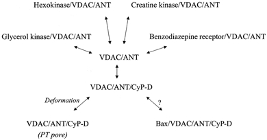
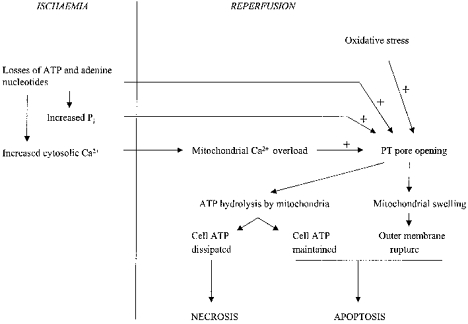
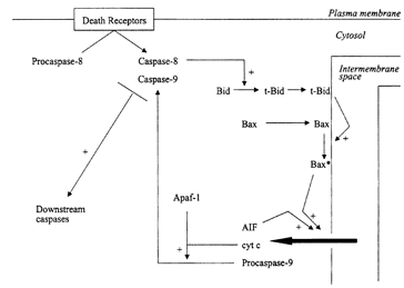
A mitochondrial complex comprising the voltage-dependent anion channel (outer membrane), the adenine nucleotide translocase (inner membrane) and cyclophilin-D (matrix) assembles at contact sites between the inner and outer membranes. Under pathological conditions associated with ischaemia and reperfusion the junctional complex 'deforms' into the permeability transition (PT) pore, which can open transiently, allowing free permeation of low Mr solutes across the inner membrane. This may be a critical step in the pathogenesis of lethal cell injury in ischaemia and reperfusion. Moreover, it is argued, the degree of pore opening may be an important determinant of the relative extent of apoptosis and necrosis under these conditions. In addition, mitochondria are the major sites of action of Bax and other apoptotic regulatory proteins of the Bcl-2 family. These proteins control a mitochondrial amplificatory loop in the apoptotic signalling pathway in which cytochrome c and other apoptogenic proteins of the mitochondrial intermembrane space are released into the cytosol. There are indications that the junctional complex, or components of it, may also mediate the action of Bax, but in a way that does not involve PT pore formation.
Figures
References
-
- Andreeva L, Tanveer A, Crompton M. Evidence for the involvement of a mitochondrial cyclosporin A binding protein in the Ca-activated inner membrane pore of heart mitochondria. European Journal of Biochemistry. 1995;230:1125–1132. - PubMed
-
- Ankarcrona M, Dypbukt JM, Bonfoco E, Zhivotovsky B, Orrenius S, Lipton SA, Nicotera P. Glutamate-induced neuronal death: a succession of necrosis or apoptosis depending on mitochondrial function. Neuron. 1995;15:961–973. - PubMed
-
- Anversa P, Cheng W, Liu Y, Leri A, Redaelli G, Kajstura J. Apoptosis and myocardial infarction. Basic Research in Cardiolology. 1998;93(suppl 3):8–12. - PubMed
Publication types
MeSH terms
Substances
LinkOut - more resources
Full Text Sources
Research Materials