Review
. 2000 Nov 15;529 Pt 1(Pt 1):23-36.
doi: 10.1111/j.1469-7793.2000.00023.x.

Pathophysiological and protective roles of mitochondrial ion channels

Affiliations
Review

Pathophysiological and protective roles of mitochondrial ion channels

B O'Rourke. J Physiol. .

Abstract

Mitochondria possess a highly permeable outer membrane and an inner membrane that was originally thought to be relatively impermeable to ions to prevent dissipation of the electrochemical gradient for protons. Although recent evidence has revealed a rich diversity of ion channels in both membranes, the purpose of these channels remains incompletely determined. Pores in the outer membrane are fundamental participants in apoptotic cell death, and this process may also involve permeability transition pores on the inner membrane. Novel functions are now being assigned to other ion channels of the inner membrane. Examples include protection against ischaemic injury by mitochondrial KATP channels and the contribution of inner membrane anion channels to spontaneous mitochondrial oscillations in cardiac myocytes. The central role of mitochondria in both the normal function of the cell and in its demise makes these channels prime targets for future research and drug development.


Figures

Figure 1
Figure 1. Mitochondrial redox and membrane potentials
The redox state of the cell depends on the rates of production and oxidation of reducing equivalents in the cytoplasm and in the mitochondrial matrix. FAD-linked dehydrogenases in the matrix including pyruvate dehydrogenase (PDH), α-ketoglutarate dehydrogenase (α-KGDH) and succinate dehydrogenase (SDH) fluoresce green (excitation 480 nm/emission peak ≈520 nm) when oxidized. These flavoproteins are in equilibrium with the mitochondrial NADH/NAD+ redox couple. NADH is also fluorescent, emitting blue light (peak ≈450 nm) with ultraviolet excitation (350 nm). Mitochondrial inner membrane potential (ΔΨ) can be assessed by the distribution of the lipophilic cations like tetramethylrhodamine ethyl ester (TMRE), which emits red fluorescence (excitation 540 nm/emission 605 nm). The opening of an inner membrane ion channel (e.g. mitoKATP) leads to acceleration of NADH oxidation by the electron transport chain and partial dissipation of the proton gradient through activation of K+-H+ exchange. A net change in ΔΨ or redox potential occurs if NADH production and proton pumping cannot match the increase in energy dissipation.
Figure 2
Figure 2. Regional depolarization of mitochondria
A, loss of ΔΨ in a substrate-deprived guinea-pig ventricular myocyte can be restricted to ‘superclusters’ of mitochondria, as shown for this series of images of TMRE fluorescence (100 nM TMRE loading). The timing of the images in seconds is denoted on each frame. B, rapid loss and slow recovery of TMRE fluorescence in the cluster (region ii) and unchanging signal in a neighbouring region (i; as shown in A) illustrate the independent behaviour of adjacent mitochondria. C, the full time course of multiple slow oscillations of ΔΨ in the supercluster.
Figure 3
Figure 3. Suppression of oscillations by a benzodiazepine receptor ligand
A, exposure to PK11195 (200 μm applied between dashed lines) reversibly suppressed oscillations in ΔΨ (TMRE signal) and flavoprotein redox (FP signal; thick line) as derived from time course image analysis. B, taking the first derivative of the redox signal (FP dF/dt) facilitates analysis of the frequency of transitions by eliminating slow baseline changes. Counting fast transitions that exceed a threshold (in this case 20 % of peak + dF/dt) permits quantitative determination of drug efficacy C, the number of oxidations over a period of 10 min in the presence of PK11195 was significantly suppressed (n = 12 cells) compared with before and after application.
Figure 4
Figure 4. Selective activation of mitoKATP by diazoxide in cardiomyocytes
A, diazoxide causes reversible and reproducible oxidation of mitochondrial flavoproteins (Diazo; 100 μm). The fluorescence signal can be calibrated by maximally oxidizing (with the uncoupler 2,4-dinitrophenol; DNP) and reducing the mitochondria (with the cytochrome oxidase inhibitor cyanide; CN). B, in the same experiment, diazoxide did not activate sarcolemmal KATP current. Severe metabolic inhibition with DNP or CN activated sarcolemmal KATP, confirming the presence of these channels. From Liu et al. (1998).
Figure 5
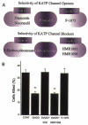
Figure 5. Summary of the selectivity of KATP channel openers and blockers
A, based on studies of intact myocytes using methods similar to that shown in Fig. 4, the selective mitoKATP agonists diazoxide and nicorandil have been identified, while pinacidil activates both mitochondrial and surface isoforms. The pinacidil derivative P-1075 selectively activates sarcolemmal KATP in intact myocytes. B, cellular protection against simulated ischaemia is conferred by diazoxide, but not P-1075, and is blocked by 5-HD, but not HMR-1098, thus supporting a mechanism involving mitoKATP rather than sarcKATP.

Similar articles

Cited by

References

    1. Amchenkova AA, Bakeeva LE, Chentsov YS, Skulachev VP, Zorov DB. Coupling membranes as energy-transmitting cables. I. Filamentous mitochondria in fibroblasts and mitochondrial clusters in cardiomyocytes. Journal of Cell Biology. 1988;107:481–495. - PMC - PubMed
    1. Antonenko YN, Kinnally KW, Tedeschi H. Identification of anion and cation pathways in the inner mitochondrial membrane by patch clamping of mouse liver mitoplasts. Journal of Membrane Biology. 1991;124:151–158. - PubMed
    1. Auchampach JA, Grover GJ, Gross GJ. Blockade of ischaemic preconditioning in dogs by the novel ATP dependent potassium channel antagonist sodium 5-hydroxydecanoate. Cardiovascular Research. 1992;26:1054–1062. - PubMed
    1. Ballarin C, Sorgato MC. An electrophysiological study of yeast mitochondria. Evidence for two inner membrane anion channels sensitive to ATP. Journal of Biological Chemistry. 1995;270:19262–19268. - PubMed
    1. Beavis AD. Properties of the inner membrane anion channel in intact mitochondria. Journal of Bioenergetics and Biomembranes. 1992;24:77–90. - PubMed

Publication types

LinkOut - more resources