

Mitochondrial signals in glucose-stimulated insulin secretion in the beta cell
- PMID: 11080250
- PMCID: PMC2270172
- DOI: 10.1111/j.1469-7793.2000.00049.x
Mitochondrial signals in glucose-stimulated insulin secretion in the beta cell
Abstract
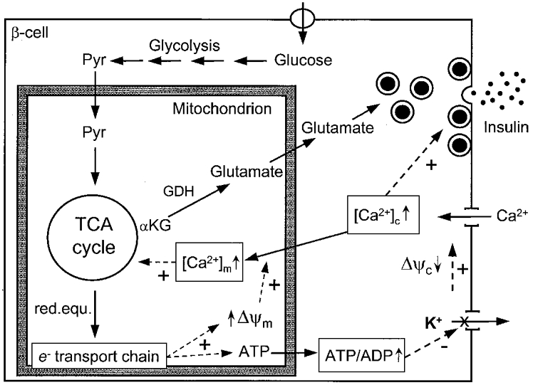
Glucose-induced insulin secretion is determined by signals generated in the mitochondria. The elevation of ATP is necessary for the membrane-dependent increase in cytosolic Ca2+, the main trigger of insulin exocytosis. Beta cells depleted of mitochondrial DNA fail to respond to glucose while still secreting insulin in response to membrane depolarisation. This cell model resembles the situation of defective insulin secretion in patients with mitochondrial diabetes. On the other hand, infants with activating mutations in the mitochondrial enzyme glutamate dehydrogenase are characterised by hyperinsulinism and hypoglycaemia. We have recently proposed that glutamate, generated by this enzyme, participates in insulin secretion as a glucose-derived metabolic messenger. In this model, glutamate acts downstream of the mitochondria by sensitising the exocytotic process to Ca2+. The evidence in favour of such a role for glutamate is discussed in the present review.
Figures
Similar articles
-
Mitochondrial glutamate acts as a messenger in glucose-induced insulin exocytosis.Nature. 1999 Dec 9;402(6762):685-9. doi: 10.1038/45280. Nature. 1999. PMID: 10604477
-
Beta-cell mitochondria in the regulation of insulin secretion: a new culprit in type II diabetes.Diabetologia. 2000 Mar;43(3):265-77. doi: 10.1007/s001250050044. Diabetologia. 2000. PMID: 10768087 Review.
-
Mitochondria as the conductor of metabolic signals for insulin exocytosis in pancreatic beta-cells.Cell Mol Life Sci. 2002 Nov;59(11):1803-18. doi: 10.1007/pl00012507. Cell Mol Life Sci. 2002. PMID: 12530515 Free PMC article. Review.
-
Modulation of glutamate generation in mitochondria affects hormone secretion in INS-1E beta cells.IUBMB Life. 2000 Jul;50(1):27-31. doi: 10.1080/15216540050176557. IUBMB Life. 2000. PMID: 11087117
-
Beta-cell mitochondria and insulin secretion: messenger role of nucleotides and metabolites.Diabetes. 2002 Feb;51 Suppl 1:S37-42. doi: 10.2337/diabetes.51.2007.s37. Diabetes. 2002. PMID: 11815456 Review.
Cited by
-
RyR channels and glucose-regulated pancreatic beta-cells.Eur Biophys J. 2008 Jul;37(6):773-82. doi: 10.1007/s00249-008-0269-0. Epub 2008 Feb 1. Eur Biophys J. 2008. PMID: 18239912
-
The mitochondrial transporter family (SLC25): physiological and pathological implications.Pflugers Arch. 2004 Feb;447(5):689-709. doi: 10.1007/s00424-003-1099-7. Epub 2003 Nov 4. Pflugers Arch. 2004. PMID: 14598172 Review.
-
Analysis of Non-Human Primate Pancreatic Islet Oxygen Consumption.J Vis Exp. 2019 Dec 18;(154):10.3791/60696. doi: 10.3791/60696. J Vis Exp. 2019. PMID: 31904022 Free PMC article.
-
Dysfunctions, molecular mechanisms, and therapeutic strategies of pancreatic β-cells in diabetes.Apoptosis. 2023 Aug;28(7-8):958-976. doi: 10.1007/s10495-023-01854-0. Epub 2023 Jun 5. Apoptosis. 2023. PMID: 37273039 Review.
-
Fumarate Hydratase Deletion in Pancreatic β Cells Leads to Progressive Diabetes.Cell Rep. 2017 Sep 26;20(13):3135-3148. doi: 10.1016/j.celrep.2017.08.093. Cell Rep. 2017. PMID: 28954230 Free PMC article.
References
-
- Ahren B. Autonomic regulation of islet hormone secretion-implications for health and disease. Diabetologia. 2000;43:393–410. - PubMed
-
- Antinozzi PA, Segall L, Prentki M, McGarry JD, Newgard CB. Molecular or pharmacologic perturbation of the link between glucose and lipid metabolism is without effect on glucose-stimulated insulin secretion. A re-evaluation of the long-chain acyl-CoA hypothesis. Journal of Biological Chemistry. 1998;273:16146–16154. - PubMed
-
- Bellocchio EE, Reimer RJ, Fremeau RT, Jr, Edwards RH. Uptake of glutamate into synaptic vesicles by an inorganic phosphate transporter. Science. 2000;289:957–960. - PubMed
-
- Bergsten P. Slow and fast oscillations of cytoplasmic Ca2+ in pancreatic islets correspond to pulsatile insulin release. American Journal of Physiology. 1995;268:E282–287. - PubMed
Publication types
MeSH terms
Substances
LinkOut - more resources
Full Text Sources
Medical
Miscellaneous