Review
. 2000 Nov 15;529 Pt 1(Pt 1):69-81.
doi: 10.1111/j.1469-7793.2000.00069.x.

The machinery of local Ca2+ signalling between sarco-endoplasmic reticulum and mitochondria

Affiliations
Review

The machinery of local Ca2+ signalling between sarco-endoplasmic reticulum and mitochondria

G Hajnóczky et al. J Physiol. .

Abstract

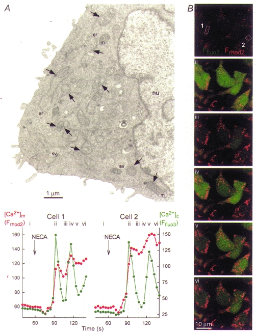
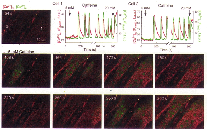
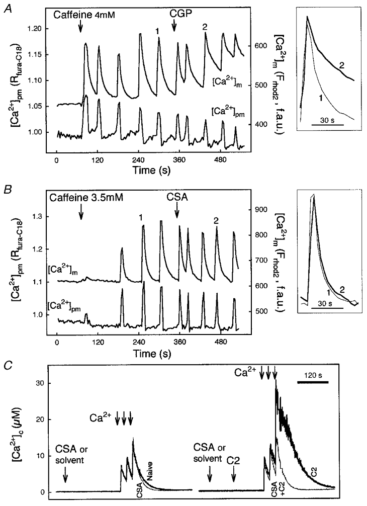
Growing evidence suggests that propagation of cytosolic [Ca2+] ([Ca2+]c) spikes and oscillations to the mitochondria is important for the control of fundamental cellular functions. Delivery of [Ca2+]c spikes to the mitochondria may utilize activation of the mitochondrial Ca2+ uptake sites by the large local [Ca2+]c rise occurring in the vicinity of activated sarco-endoplasmic reticulum (SR/ER) Ca2+ release channels. Although direct measurement of the local [Ca2+]c sensed by the mitochondria has been difficult, recent studies shed some light onto the molecular mechanism of local Ca2+ communication between SR/ER and mitochondria. Subdomains of the SR/ER are in close contact with mitochondria and display a concentration of Ca2+ release sites, providing the conditions for an effective delivery of released Ca2+ to the mitochondrial targets. Furthermore, many functional properties of the signalling between SR/ER Ca2+ release sites and mitochondrial Ca2+ uptake sites, including transient microdomains of high [Ca2+], saturation of mitochondrial Ca2+ uptake sites by released Ca2+, connection of multiple release sites to each uptake site and quantal transmission, are analogous to the features of the coupling between neurotransmitter release sites and postsynaptic receptors in synaptic transmission. As such, Ca2+ signal transmission between SR/ER and mitochondria may utilize discrete communication sites and a closely related functional architecture to that used for synaptic signal propagation between cells.


Figures

Figure 1
Figure 1. Physical and functional coupling between ER and mitochondria in RBL-2H3 mast cells
A, electron micrograph showing a thin section (80 nm) of a RBL-2H3 mast cell. Organelles are marked as follows: nu, nucleus; er, endoplasmic reticulum; m, mitochondria; sv, secretory vesicles. Arrows point to ER-mitochondrial junctions. Note that only a few examples of ER, mitochondria and ER-mitochondrial junctions are marked. B, simultaneous confocal imaging of IP3R-driven [Ca2+]c and [Ca2+]m signals in RBL-2H3 cells. Cells were loaded first with rhod-2 AM ([Ca2+]m; 3 μm for 50 min at 37 °C) and subsequently with fluo-3 AM ([Ca2+]c; 5 μm for 25 min at room temperature). Evidence that rhod-2 measured [Ca2+]m in RBL-2H3 cells was provided by morphological and pharmacological studies described in Csordás et al. 1999. Fluorescence intensity of fluo-3 and rhod-2 reflecting [Ca2+]c and [Ca2+]m are depicted on linear green and red scales, respectively. Confocal image time series shows the spatiotemporal pattern of [Ca2+] responses evoked by a phospholipase C-linked adenosine receptor agonist, 5′-(N-ethyl)carboxamidoadenosine (NECA, 50 μm). Graphs show corresponding traces of [Ca2+]c and [Ca2+]m (fluo-3 and rhod-2 signals, respectively, expressed as fluorescence arbitrary units) calculated for the regions marked by boxes on image i.
Figure 2
Figure 2. Coordination of RyR-driven [Ca2+]c and [Ca2+]m oscillations and waves in permeabilized H9c2 cardiac myotubes
Simultaneous confocal imaging of [Ca2+]c and [Ca2+]m carried out using fluo-3 and compartmentalized rhod-2, respectively. Cells were loaded first with rhod-2 AM (4 μm for 50 min at 37 °C) and after permeabilization, fluo-3 FA (10 μm) was added to the intracellular medium. Measurement of [Ca2+]m with rhod-2 in permeabilized myotubes has been described in Szalai et al. 2000. Fluorescence intensity of fluo-3 and rhod-2 reflecting [Ca2+]c and [Ca2+]m are depicted on linear green and red scales, respectively. Confocal image time series shows the spatiotemporal pattern of [Ca2+] responses evoked by a RyR activator, caffeine. [Ca2+]c and [Ca2+]m spikes propagated through the myotubes as waves (middle row of images shows the first wave, whereas the lower row of images shows the second wave). Graphs show corresponding traces of [Ca2+]c and [Ca2+]m calculated for the regions marked by boxes on the image in the upper row. f.a.u., fluorescence arbitrary units.
Figure 3
Figure 3. Ca2+ release from mitochondria in naive cells and in cells exposed to C2-ceramide
A and B, time courses of perimembrane [Ca2+] ([Ca2+]pm) and [Ca2+]m, and responses evoked by caffeine in two individual rhod-2-loaded permeabilized myotubes. [Ca2+]pm was monitored using fura-C18. Effect of CGP 37157 (CGP, 10 μm; A) and cyclosporin A (CSA, 1 μm; B) on caffeine-induced [Ca2+] oscillations. Insets: [Ca2+]m spikes recorded prior to and after addition of the drug are shown by synchronizing the rising phase. Reproduced with permission from Szalai et al. (2000). C, effect of CSA on mitochondrial Ca2+ sequestration evoked by Ca2+ pulsing (3 pulses, 25 μm CaCl2 each) in suspensions of naive (left) and C2-ceramide-pretreated (C2; 40 μm for 3 min; right) permeabilized HepG2 cells. In contrast to the imaging studies, intracellular Ca2+ stores were able to control global medium [Ca2+] ([Ca2+]c) in the cell suspension studies, since the ratio of cell mass to bath volume was > 20 times larger than that in the imaging experiments. Measurements of [Ca2+]c were carried out using fura-2 FF/free acid added to the intracellular medium as described in Szalai et al. 1999. The Kd value of 3 μm was determined in intracellular medium (G. Csordás & G. Hajnóczky, manuscript in preparation) and used to translate the fura-2 FF fluorescence ratios to [Ca2+] concentrations.

References

    1. Altschuld RA, Hohl CM, Castillo LC, Garleb AA, Starling RC, Brierley GP. Cyclosporin inhibits mitochondrial calcium efflux in isolated adult rat ventricular cardiomyocytes. American Journal of Physiology. 1992;262:1699–1704. - PubMed
    1. Babcock DF, Herrington J, Goodwin PC, Park YB, Hille B. Mitochondrial participation in the intracellular Ca2+ network. Journal of Cell Biology. 1997;136:833–844. - PMC - PubMed
    1. Babcock DF, Hille B. Mitochondrial oversight of cellular Ca2+ signalling. Current Opinions in Neurobiology. 1998;8:398–404. - PubMed
    1. Bassani RA, Bassani JW, Bers DM. Mitochondrial and sarcolemmal Ca2+ transport reduce [Ca2+]i during caffeine contractures in rabbit cardiac myocytes. The Journal of Physiology. 1992;453:591–608. - PMC - PubMed
    1. Bereiter-Hahn J, Voth M. Dynamics of mitochondria in living cells: shape changes, dislocations, fusion, and fission of mitochondria. Microscopy Research and Technique. 1994;27:198–219. - PubMed

Publication types

LinkOut - more resources