. 2000 Nov;124(3):1049-58.
doi: 10.1104/pp.124.3.1049.

Local and systemic induction of two defense-related subtilisin-like protease promoters in transgenic Arabidopsis plants. Luciferin induction of PR gene expression

Affiliations

Local and systemic induction of two defense-related subtilisin-like protease promoters in transgenic Arabidopsis plants. Luciferin induction of PR gene expression

L Jordá et al. Plant Physiol. 2000 Nov.

Abstract

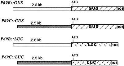
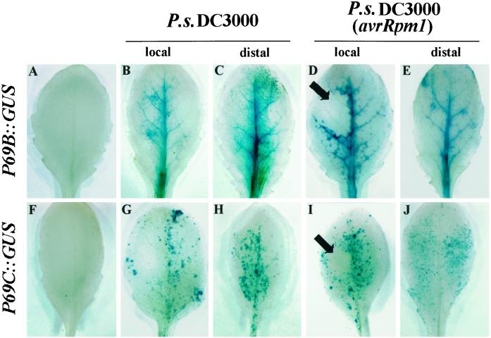
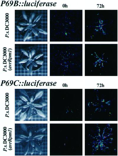
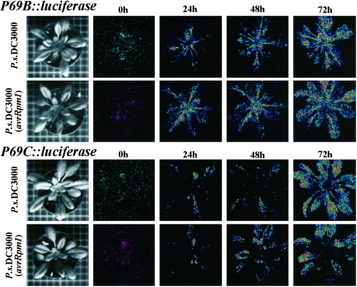
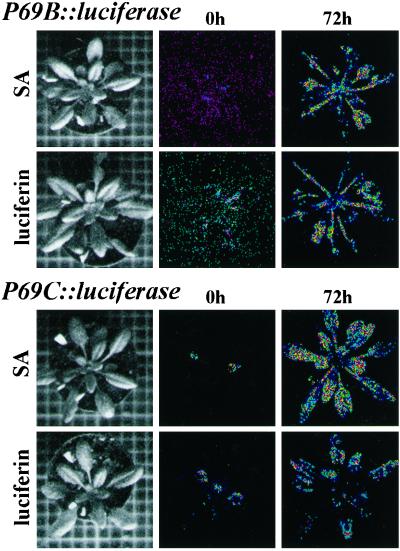
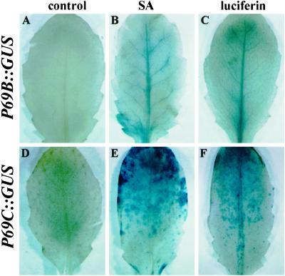
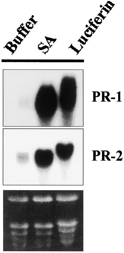
Following a pathogenic attack, plants are able to mount a defense response with the coordinated activation of a battery of defense-related genes. In this study we have characterized the mode of expression of the P69B and P69C genes from tomato (Lycopersicon esculentum Mill.), which encodes two closely related subtilisin-like proteases associated with the defense response. We have compared the mode of gene regulation in heterologous transgenic Arabidopsis plants harboring promoter-beta-glucuronidase (GUS) and promoter-luciferase (LUC) gene fusions for these two genes. These studies revealed that the P69B and P69C promoters are induced by salicylic acid as well as during the course of both a compatible and an incompatible interaction with Pseudomonas syringae. Furthermore, P69B and P69C expression takes place in both the local and the distal (noninoculated) leaves upon inoculation with bacteria but following different and unique tissue-specific patterns of expression that are also different to that described for most other classical PR genes. Also, we report that luciferin, the substrate for the reporter luciferase (LUC) gene, is able to activate expression of PR genes, and this may pose a problem when using this gene reporter system in studies related to plant defense.


Figures

Figure 1
Figure 1
Schematic representation of the different P69::GUS and P69::LUC gene fusions. The diagonally striped boxes represent the GUS or LUC genes. The white box at the right represents the 3′-region of the nopaline synthase gene. The length of each of the promoter regions is shown above each construct in kilobase pairs. The ATG codon represents the first translation initiation codon that resides in the reporter gene.
Figure 2
Figure 2
GUS staining patterns in rosette leaves of transgenic Arabidopsis (Col-0) plants carrying the P69B::GUS and P69C::GUS transgenes. Top, GUS staining pattern in leaves from P69B::GUS transgenic plants. Bottom, GUS staining pattern in leaves from P69C::GUS. A and F, GUS staining pattern in leaves from noninoculated plants. B and G, GUS staining pattern in leaves inoculated with P.s. DC3000. C and H, GUS staining pattern in distal (noninoculated) leaves from plants inoculated with P.s. DC3000. D and I, GUS staining pattern in leaves inoculated with P.s. DC3000 carrying the avirulent Rpm1 gene. E and J, GUS staining pattern in distal (noninoculated) leaves from plants inoculated with P.s. DC3000 carrying the avirulent Rpm1 gene. Leaves were analyzed 72 h after inoculation. The characteristic HR responses elicited in the inoculated leaves with the incompatible bacteria are indicated with arrows. The experiments were repeated with plants from at least three different transgenic lines for each construct and in all cases render similar results.
Figure 3
Figure 3
Monitoring of light emissions in entire P69B::LUC and P69C::LUC transgenic Arabidopsis plants by low-light video image analysis following inoculation with compatible and incompatible bacteria. Left, Standard photographs of the type of plants used in these experiments. Middle, Luminescence from plants at the time of inoculation (0 h) with either P.s. DC3000 or P.s. DC3000 (avrRpm1). Right, Luminescence from plants at 72-h postinoculation with either P.s. DC3000 or P.s. DC3000 (avrRpm1). At the times indicated, plants were sprayed once with 1 mm luciferin, and images were obtained immediately after 10 min of photon collection. The plants shown in each of the panels are different plants but derived from the same homozygous transgenic lines. The experiments were repeated with plants from at least three different transgenic lines for each construct and in all cases render similar results.
Figure 4
Figure 4
Monitoring of light emissions by low-light video image analysis in a single transgenic Arabidopsis plant during the course of a compatible or an incompatible interaction. Two leaves from each plant were inoculated with either P.s. DC3000 or P.s. DC3000 (AvrRpm1) and at each time point the plant was taken from the growth chamber, sprayed with 1 mm luciferin, and immediately the photon collection was performed for 10 min. This process was repeated in the same plant at 0, 24, 48, and 72 h postinoculation with the bacteria. The left column of pictures shows a standard photograph of the single plant used in each experiment. Top, Two plants derived from the same P69B::LUC transgenic line. Bottom, Two plants derived from the same P69C::LUC transgenic line. The experiments were repeated with plants from at least three different transgenic lines for each construct and in all cases render similar results.
Figure 5
Figure 5
Effect of exogenous application of SA and luciferin on the activation of P69B::LUC and P69C::LUC gene expression. P69B::LUC transgenic Arabidopsis plants (top) and P69C::LUC transgenic Arabidopsis plants (bottom) were sprayed three times (at 24-h intervals) with a solution containing 0.5 mm SA or 1 mm luciferin. The same plants were monitored for light emissions by low-light video image at 0 or 72 h after receiving the first chemical treatment. Images were obtained after 10 min of photon collection.
Figure 6
Figure 6
Effect of SA and luciferin on the activation of P69B::GUS and P69C::GUS gene expression. P69B::GUS transgenic Arabidopsis plants (top) and P69C::GUS transgenic Arabidopsis plants (bottom) were sprayed three times (at 24-h intervals) with a solution containing 0.5 mm SA or 1 mm luciferin, and GUS expression was detected by histochemical staining of leaves with X-gluc. A and D, GUS staining pattern before treatment. B and E, GUS staining pattern at 72 h after SA treatments. C and F, GUS staining pattern at 72 h after luciferin treatments.
Figure 7
Figure 7
Northern-blot analyses of endogenous Arabidopsis PR-1 and PR-2 gene expression at 48 h following treatment of plants with SA and luciferin.

Similar articles

Cited by

References

    1. Agrios GN. Plant Pathology. London: Academic Press; 1988.
    1. Alonso E, de Carvalho Niebel E, Obregón P, Gheysen G, Inzé D, Van Montagu M, Castresana C. Differential in vitro DNA binding activity to a promoter element of the gn1 β-1,3-glucanase gene in hypersensitively reacting tobacco plants. Plant J. 1995;7:309–320. - PubMed
    1. Bechtold N, Ellis J, Pelletier G. In planta Agrobacterium mediated gene transfer by infiltration of adult Arabidopsis thaliana plants. CR Acad Sci Paris Life Sci. 1993;316:1194–1199.
    1. Brederode FT, Linthorst HJM, Bol JF. Differential induction of acquired resistance and PR gene expression in tobacco by virus infection, ethephon treatment, UV light and wounding. Plant Mol Biol. 1991;17:1117–1125. - PubMed
    1. Broglie K, Chet I, Holliday M, Cressman R, Riddle P, Knowlton S, Mauvais CJ, Broglie R. Transgenic plants with enhanced resistance to the fungal pathogen Rhizoctonia solani. Science. 1991;254:1194–1197. - PubMed

Publication types

MeSH terms