

Isolation of Medicago truncatula mutants defective in calcium oxalate crystal formation
- PMID: 11080287
- PMCID: PMC59209
- DOI: 10.1104/pp.124.3.1097
Isolation of Medicago truncatula mutants defective in calcium oxalate crystal formation
Abstract
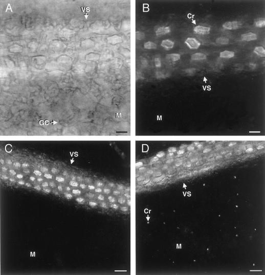
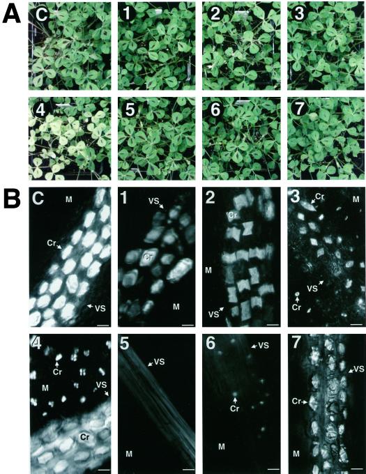
Plants accumulate crystals of calcium oxalate in a variety of shapes, sizes, amounts, and spatial locations. How and why many plants form crystals of calcium oxalate remain largely unknown. To gain insight into the regulatory mechanisms of crystal formation and function, we have initiated a mutant screen to identify the genetic determinants. Leaves from a chemically mutagenized Medicago truncatula population were visually screened for alterations in calcium oxalate crystal formation. Seven different classes of calcium oxalate defective mutants were identified that exhibited alterations in crystal nucleation, morphology, distribution and/or amount. Genetic analysis suggested that crystal formation is a complex process involving more than seven loci. Phenotypic analysis of a mutant that lacks crystals, cod 5, did not reveal any difference in plant growth and development compared with controls. This finding brings into question the hypothesized roles of calcium oxalate formation in supporting tissue structure and in regulating excess tissue calcium.
Figures
Similar articles
-
Calcium oxalate crystal morphology mutants from Medicago truncatula.Planta. 2002 Jul;215(3):380-6. doi: 10.1007/s00425-002-0759-8. Epub 2002 Apr 20. Planta. 2002. PMID: 12111218
-
Isolated Medicago truncatula mutants with increased calcium oxalate crystal accumulation have decreased ascorbic acid levels.Plant Physiol Biochem. 2007 Mar-Apr;45(3-4):216-20. doi: 10.1016/j.plaphy.2007.01.013. Epub 2007 Feb 4. Plant Physiol Biochem. 2007. PMID: 17400466
-
Influence of calcium oxalate crystal accumulation on the calcium content of seeds from Medicago truncatula.Plant Sci. 2012 Apr;185-186:246-9. doi: 10.1016/j.plantsci.2011.11.004. Epub 2011 Nov 9. Plant Sci. 2012. PMID: 22325887
-
Calcium oxalate in plants: formation and function.Annu Rev Plant Biol. 2005;56:41-71. doi: 10.1146/annurev.arplant.56.032604.144106. Annu Rev Plant Biol. 2005. PMID: 15862089 Review.
-
Oxalate binding proteins in calcium oxalate nephrolithiasis.Urol Res. 2003 Aug;31(4):242-56. doi: 10.1007/s00240-003-0316-3. Epub 2003 Jul 11. Urol Res. 2003. PMID: 12856168 Review.
Cited by
-
Plant calcium content: ready to remodel.Nutrients. 2012 Aug;4(8):1120-36. doi: 10.3390/nu4081120. Epub 2012 Aug 21. Nutrients. 2012. PMID: 23016135 Free PMC article.
-
Medicago truncatula mutants demonstrate the role of plant calcium oxalate crystals as an effective defense against chewing insects.Plant Physiol. 2006 May;141(1):188-95. doi: 10.1104/pp.106.076737. Epub 2006 Mar 2. Plant Physiol. 2006. PMID: 16514014 Free PMC article.
-
Isolation of a crystal matrix protein associated with calcium oxalate precipitation in vacuoles of specialized cells.Plant Physiol. 2003 Oct;133(2):549-59. doi: 10.1104/pp.103.023556. Plant Physiol. 2003. PMID: 14555781 Free PMC article.
-
Increased calcium bioavailability in mice fed genetically engineered plants lacking calcium oxalate.Plant Mol Biol. 2007 Jul;64(5):613-8. doi: 10.1007/s11103-007-9180-9. Epub 2007 May 20. Plant Mol Biol. 2007. PMID: 17514431
-
An Assessment of Engineered Calcium Oxalate Crystal Formation on Plant Growth and Development as a Step toward Evaluating Its Use to Enhance Plant Defense.PLoS One. 2015 Oct 30;10(10):e0141982. doi: 10.1371/journal.pone.0141982. eCollection 2015. PLoS One. 2015. PMID: 26517544 Free PMC article.
References
-
- Arnott HJ, Pautard FGE. Calcification in plants. In: Schraer H, editor. Biological Calcification: Cellular and Molecular Aspects. New York: Appleton-Century-Crofts; 1970. pp. 375–446.
-
- Arnott HJ, Webb MA. Twin crystals of calcium oxalate in the seed coat of the kidney bean. Protoplasma. 1983;114:23–34.
-
- Baggio B, Gambaro G. Cellular abnormalities of oxalate transport in nephrolithiasis. In: Khan SR, editor. Calcium Oxalate in Biological Systems. Boca Raton, FL: CRC Press; 1995. p. 207.
-
- Borchert R. Calcium-induced patterns of calcium-oxalate crystals in isolated leaflets of Gleditsia triacanthos L. and Albizia julibrissin Durazz. Planta. 1985;165:301–310. - PubMed
-
- Borchert R. Calcium acetate induces calcium uptake and formation of calcium-oxalate crystals in isolated leaflets of Gleditsia tracanthos L. Planta. 1986;168:571–578. - PubMed
Publication types
MeSH terms
Substances
LinkOut - more resources
Full Text Sources
