. 2000 Nov;124(3):1229-38.
doi: 10.1104/pp.124.3.1229.

Overexpression of auxin-binding protein enhances the sensitivity of guard cells to auxin

Affiliations

Overexpression of auxin-binding protein enhances the sensitivity of guard cells to auxin

J M Bauly et al. Plant Physiol. 2000 Nov.

Abstract

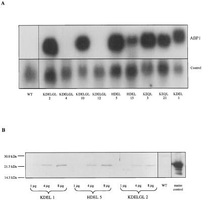
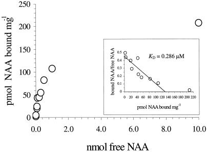
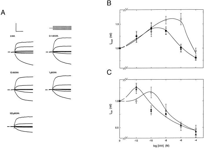
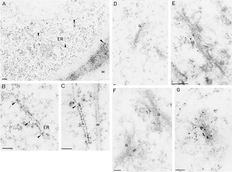
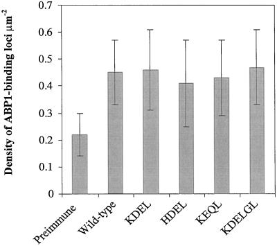
To explore the role of auxin-binding protein (ABP1) in planta, a number of transgenic tobacco (Nicotiana tabacum) lines were generated. The wild-type KDEL endoplasmic reticulum targeting signal was mutated to HDEL, another common retention sequence in plants, and to KEQL or KDELGL to compromise its activity. The auxin-binding kinetics of these forms of ABP1 were found to be similar to those of ABP1 purified from maize (Zea mays). To test for a physiological response mediated by auxin, intact guard cells of the transgenic plants were impaled with double-barreled microelectrodes, and auxin-dependent changes in K(+) currents were recorded under voltage clamp. Exogenous auxin affected inwardly and outwardly rectifying K(+) currents in a dose-dependent manner. Auxin sensitivity was markedly enhanced in all plants overexpressing ABP1, irrespective of the form present. Immunogold electron microscopy was used to investigate the localization of ABP1 in the transgenic plants. All forms were detected in the endoplasmic reticulum and the KEQL and KDELGL forms passed further across the Golgi stacks than KDEL and HDEL forms. However, neither electron microscopy nor silver-enhanced immunogold epipolarization microscopy revealed differences in cell surface ABP1 abundance for any of the plants, including control plants, which indicated that overexpression of ABP1 alone was sufficient to confer increased sensitivity to added auxin. Jones et al. ([1998] Science 282: 1114-1117) found increased cell expansion in transgenic plants overexpressing wild-type ABP1. Single cell recordings extend this observation, with the demonstration that the auxin sensitivity of guard cell K(+) currents is mediated, at least in part, by ABP1.


Figures

Figure 1
Figure 1
Expression of the ABP1 transgenes. A, Northern blot showing maize ABP1 mRNA accumulation in primary transformants. Blots were hybridized sequentially to a maize ABP1 probe (ABP1) and the constitutively expressed pCNT 6 (Memelink et al., 1987; control). The larger size of KDEL-ABP1 mRNA compared with the mutated ABP1 mRNAs is due to the (intentional) loss of some 3′-untranslated region during the in vitro mutagenesis schedule. The maize ABP1 probe did not detect wild-type tobacco ABP1 in untransformed plants (WT). B, Representative immunoblot showing expression of ABP1 in selected transgenic plants. Total microsomal protein loading ranged from 1 to 8 μg, as indicated, for the transgenic lines and 20 μg for microsomal protein from a wild-type (untransformed) tobacco plant (WT). ABP1 (250 ng) from maize microsomes was used as a positive control (maize control).
Figure 2
Figure 2
Auxin-binding activity. All forms of maize ABP1 displayed similar binding kinetics regardless of the C-terminal sequence. A representative plot for the KDELGL form, adjusted for non-saturable background, is shown. Inset, Estimates of KD were made by Scatchard analysis.
Figure 3
Figure 3
Potassium currents from voltage-clamped guard cells. A, K+ current from one guard cell of a tobacco plant carrying the KDEL form of the ABP1 transgene. Data recorded in 5 mm Ca2+-MES, pH 6.1, with 10 mm KCl and IAA added at the concentrations indicated. All measurements shown were carried out after current responses reached steady state with respect to auxin. Clamp cycle (above, right): −100 mV conditioning voltage followed by steps (eight cycles) to voltages of +30 to −200 mV. Scale (above, left): vertical 100 μA cm−2 or 600 mV; horizontal, 2 s. Current zero indicated on left by each set of traces. B, Response of IK,in and IK,out (C) currents to IAA concentrations between 1 pM and 100 μm. Data were pooled as indicated in the text for wild-type plus vector control (o), and ABP1 transgenics. (●), KDEL/HDEL ABP1; (▴), KEQL/KDELGL ABP1. Points represent means from 12 to 15 independent experiments, in each case normalized to their respective controls prior to auxin addition. IK,out values were taken from currents recorded at +20 mV. IK,in values were taken from currents recorded at −200 mV.
Figure 4
Figure 4
Time course of expression of ABP1 in Spodoptera frugiperda cells. A, Representative immunoblot for expression of KDEL-ABP1 up to 120 h postinfection. KDEL and HDEL forms of ABP1 were efficiently retained until cell lysis, which occurs between 72 and 96 h postinfection; thereafter, some ABP1 reaches the medium. B, Representative immunoblot for the expression of KEQL-ABP1. The KEQL and KDELGL forms of ABP1 were constitutively secreted. CELLS, Protein from cell lysates; MEDIUM, protein from cell culture medium.
Figure 5
Figure 5
Immunolocalization of ABP1 in transgenic tobacco. A through C, Labeling of the luminal ER was observed for all forms of ABP1. Sections from plants expressing KEQL (A) and KDEL (B and C) forms of ABP1 are shown. D, Control, the primary antibody was pre-incubated with purified ABP1; E, KDEL (and HDEL, not shown). ABP1 is confined to one side of the Golgi stack. F and G, KDELGL (and KEQL, not shown). ABP1 labeling in the Golgi extends from the cis to the trans side. W, Cell wall; ER, endoplasmic reticulum; G, Golgi stack. Scale bar = 100 nm. In each case representative micrographs are shown.
Figure 6
Figure 6
Measurements of ABP1 at the surface of tobacco protoplasts by SEIG EPOM. ABP1 was assayed by point light sources at the protoplast surface and expressed as density of ABP1 loci μm−2. Preimmune, Background density of point light sources on protoplasts from wild-type untransformed plants. For each genotype, 50 protoplasts were examined. Error bars show sd.

References

    1. Abel S, Theologis A. Early genes and auxin action. Plant Physiol. 1996;111:9–17. - PMC - PubMed
    1. Andres DA, Rhodes JD, Meisel RL, Dixon JE. Characterization of the carboxyl-terminal sequences responsible for protein retention in the endoplasmic reticulum. J Biol Chem. 1991;266:14277–14282. - PubMed
    1. Barbier-Brygoo H, Ephritikhine G, Klämbt D, Ghislain M, Guern J. Functional evidence for an auxin receptor at the plasmalemma of tobacco mesophyll protoplasts. Proc Natl Acad Sci USA. 1989;86:891–895. - PMC - PubMed
    1. Barbier-Brygoo H, Ephritikine G, Klämbt D, Maurel C, Palme K, Schell J, Guern J. Perception of the auxin signal at the plasma membrane of tobacco mesophyll protoplasts. Plant J. 1991;1:83–93. - PubMed
    1. Barbier-Brygoo H, Zimmermann S, Thomine S, White IR, Millner P, Guern J. Elementary response chains at the plasma membrane involve external ABP1 and multiple electrogenic ion transport proteins. Pant Growth Regul. 1996;18:23–28.

Publication types

MeSH terms

LinkOut - more resources