. 2000 Nov 2;408(6808):106-11.
doi: 10.1038/35040593.

Signal-dependent nuclear export of a histone deacetylase regulates muscle differentiation

Affiliations

Signal-dependent nuclear export of a histone deacetylase regulates muscle differentiation

T A McKinsey et al. Nature. .

Abstract

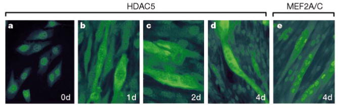
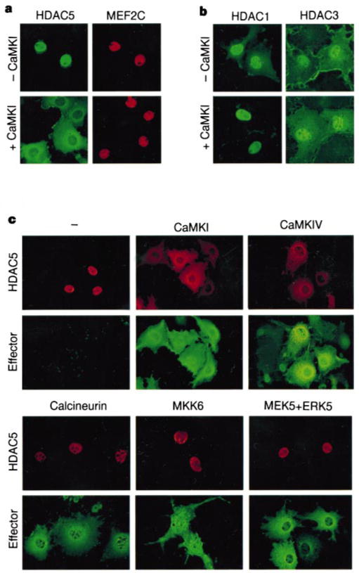
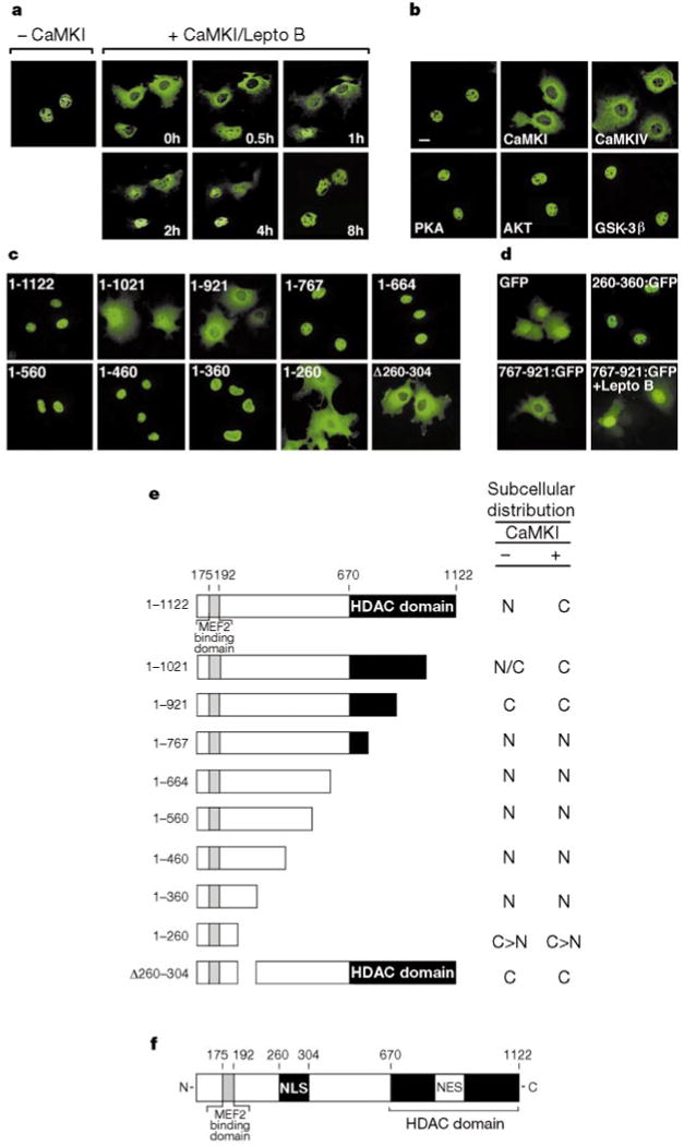
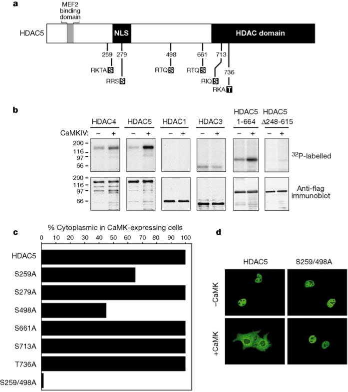
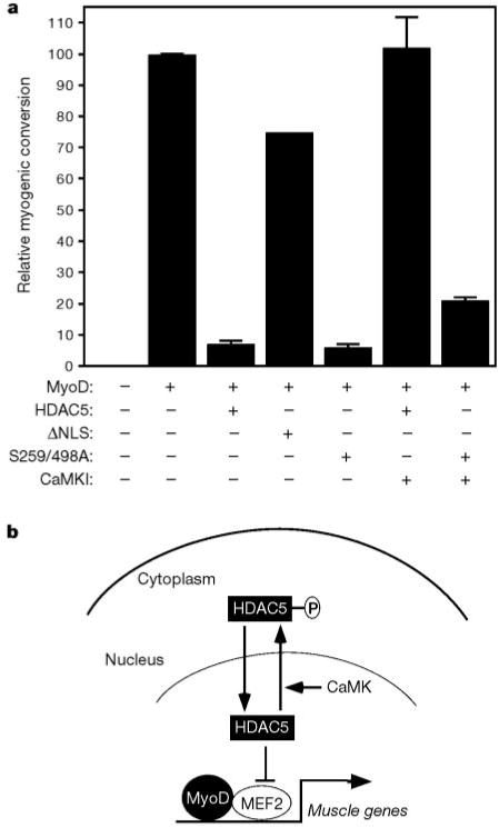
Members of the myocyte enhancer factor-2 (MEF2) family of transcription factors associate with myogenic basic helix-loop-helix transcription factors such as MyoD to activate skeletal myogenesis. MEF2 proteins also interact with the class II histone deacetylases HDAC4 and HDAC5, resulting in repression of MEF2-dependent genes. Execution of the muscle differentiation program requires release of MEF2 from repression by HDACs, which are expressed constitutively in myoblasts and myotubes. Here we show that HDAC5 shuttles from the nucleus to the cytoplasm when myoblasts are triggered to differentiate. Calcium/calmodulin-dependent protein kinase (CaMK) signalling, which stimulates myogenesis and prevents formation of MEF2-HDAC complexes, also induces nuclear export of HDAC4 and HDAC5 by phosphorylation of these transcriptional repressors. An HDAC5 mutant lacking two CaMK phosphorylation sites is resistant to CaMK-mediated nuclear export and acts as a dominant inhibitor of skeletal myogenesis, whereas a cytoplasmic HDAC5 mutant is unable to block efficiently the muscle differentiation program. Our results highlight a mechanism for transcriptional regulation through signal- and differentiation-dependent nuclear export of a chromatin-remodelling enzyme, and suggest that nucleo-cytoplasmic trafficking of HDACs is involved in the control of cellular differentiation.


Figures

Figure 1
Figure 1
Shuttling of HDAC5 from the nucleus to the cytoplasm during myogenic differentiation. Undifferentiated C2 myoblasts maintained in growth medium (a), and cells exposed to differentiation medium for one (b), two (c) or four (d) days were stained with antibodies directed against the N terminus of HDAC5 and a fluorescein-conjugated secondary antibody. We note that HDAC5 staining remains mostly nuclear in myoblasts that fail to differentiate into myotubes, whereas it is mostly cytoplasmic in adjacent myotubes. Myotubes were also stained with antiserum that recognizes both MEF2A and MEF2C (e). Images in b and c were magnified about 1.5 times relative to those in a, d and e to enable better visualization of elongated cells in the early stages of differentiation.
Figure 2
Figure 2
HDAC5 is excluded from the nucleus in cells expressing activated forms of CaMK. a, b, Cos cells were co-transfected with expression vectors encoding Flag-tagged HDAC5 and Myc-tagged MEF2C (a) or Flag-tagged HDAC1 or HDAC3 (b) in the absence or presence of a plasmid for activated CaMKI. HDACs and MEF2C were detected by indirect immunofluorescence using antibodies against the epitope tags and fluorescein-conjugated (HDACs) and rhodamine-conjugated (MEF2C) secondary antibodies. c, Cos cells were co-transfected with expression vectors for Myc-tagged HDAC5 and epitope-tagged derivatives of the indicated MEF2 activators. Cells were analysed by indirect immunofluorescence using antibodies against the epitope tags and rhodamine-conjugated (HDAC5) and fluorescein-conjugated (Effector) secondary antibodies. ERK5 staining is shown for cells co-expressing MEK5 and ERK5.
Figure 3
Figure 3
CaMK signalling stimulates nuclear export of HDAC5. a, Cos cells were transfected with an expression vector encoding HDAC5 fused to GFP in the absence or presence of a vector for activated CaMKI. GFP-positive cells were photographed before and after addition of leptomycin B (20 ng ml−1) to the medium for the indicated times. b, c, Cos cells were transfected with expression vectors for Flag-tagged HDAC5 and activated AKT, wild-type PKA, or wild-type GSK-3β (b) or Flag-tagged HDAC5 deletion mutants (c). HDAC5 was detected by indirect immunofluorescence using anti-Flag antibodies and a fluorescein-conjugated secondary antibody. d, Cos cells were transfected with expression vectors for the indicated HDAC5–GFP fusion proteins. GFP-positive cells were photographed 24 h after transfection. Leptomycin B (20 ng ml−1) was added to HDAC5(767–921)–GFP expressing cells 8 h before analysis. e, Subcellular distribution of HDAC5 mutants in the absence and presence of activated CaMK. C, cytoplasmic; N, nuclear; N/C, whole-cell; C ⟩ N, cytoplasmic greater than nuclear. f, Diagram of HDAC5.
Figure 4
Figure 4
Identification of CaMK target sites in HDAC5. a, Six consensus CaMK sites in HDAC5 that are conserved in HDAC4 are shown. b, Cos cells were transfected with expression vectors encoding Flag-tagged forms of the indicated HDAC or a Flag-tagged derivative of activated CaMKIV and Flag-tagged proteins were immunoprecipitated and incorporated into in vitro phosphorylation reactions (see Methods). Proteins were resolved by SDS–PAGE, transferred to PVDF membranes, and visualized by autoradiography (top) followed by immunoblotting with anti-Flag antibodies (bottom). c, Cos cells were co-transfected with expression vectors for Flag-tagged forms of the indicated HDAC5 point mutants and an expression plasmid for an HA-tagged derivative of activated CaMKI. Cells were analysed by double immunofluorescence using a monoclonal anti-Flag antibody and polyclonal antibodies against the HA tag. Values represent the percentage of CaMKI-expressing cells in which HDAC5 exhibited cytoplasmic staining. In d, the immunofluorescence images reveal the loss of CaMK responsiveness of the HDAC5(S259/498A) double mutant.
Figure 5
Figure 5
Regulation of myogenesis by HDAC5 nuclear export. a,10T1/2 fibroblasts were co-transfected with expression vectors for MyoD and the indicated HDAC5 protein in the absence or presence of a plasmid for activated CaMKI. Cells were transferred to differentiation medium 2 d post-transfection and stained with anti-myosin antibodies after 4d more in culture. Relative myogenic conversion represents the percentage of myosin-positive cells in HDAC transfectants relative to cells expressing MyoD alone (100 represents about 500 myosin-positive cells per 35-mm dish). Values represent the mean ± s.d. from at least two experiments. b, Model for signal-dependent regulation of myogenesis. HDAC5 blocks muscle differentiation by repressing the transcriptional activity of MEF2. CaMK phosphorylates HDAC5 and stimulates its nuclear export, freeing MEF2 to cooperate with MyoD to activate genes required for skeletal myogenesis.

Comment in

References

    1. Molkentin JD, Black BL, Martin JF, Olson EN. Cooperative activation of muscle gene expression by MEF2 and myogenic bHLH proteins. Cell. 1995;83:1125–1136. - PubMed
    1. Sparrow DB, et al. MEF-2 function is modified by a novel co-repressor, MITR. EMBO J. 1999;18:5085–5098. - PMC - PubMed
    1. Miska EA, et al. HDAC4 deacetylase associates with and represses the MEF2 transcription factor. EMBO J. 1999;18:5099–5107. - PMC - PubMed
    1. Lu J, McKinsey TA, Nicol RL, Olson EN. Signal-dependent activation of the MEF2 transcription factor by dissociation from histone deacetylases. Proc Natl Acad Sci USA. 2000;97:4070–4075. - PMC - PubMed
    1. Lu J, McKinsey TA, Zhang CL, Olson EN. Regulation of skeletal myogenesis by association of MEF2 with class II histone deacetylases. Mol Cell. 2000;6:233–244. - PubMed

Publication types

MeSH terms