

Review
doi: 10.1128/AAC.44.12.3249-3256.2000.
Aminoglycosides: perspectives on mechanisms of action and resistance and strategies to counter resistance
Affiliations
- PMID: 11083623
- PMCID: PMC90188
- DOI: 10.1128/AAC.44.12.3249-3256.2000
Item in Clipboard
Review
Aminoglycosides: perspectives on mechanisms of action and resistance and strategies to counter resistance
Antimicrob Agents Chemother.
2000 Dec.
No abstract available
Figures
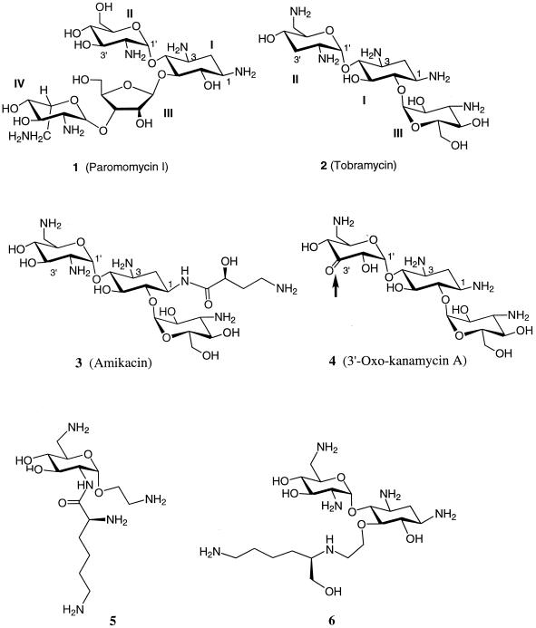
Structures of aminoglycosides.
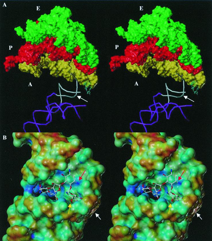
(A) Stereo view of the partial structure of the 70S rRNA complexed with three tRNA molecules (Protein Databank code, 486D [available at http://www.rcsb.rutgers.edu ]). The A-site region on the 16S rRNA is shown in white, where aminoacyl tRNA ‘A‘ (in yellow) is bound near the A-site of rRNA. Two other tRNAs, peptidyl and exit, “P” (in red) and “E” (in green), respectively, are shown as well. The backbone of the penultimate stem of the 16S rRNA molecule and the 900-loop are shown in violet. The binding site of paromomycin at the A-site is indicated by the white arrow. (B) Stereo view of the solution structure of RNA A-site template bound by paromomycin, which approximately corresponds to the A-site region on the 16S rRNA in white in panel A. The Connolly surface of the A-site RNA is rendered according to the electrostatic potential by using the MOLCAD program (Tripos, Inc., St. Louis, Mo.), and the aminoglycoside is shown in a ball-and-stick representation. The most electronegative potential is rendered in blue, and the most electropositive potential is rendered in red on the surface; all other colors show the potentials between blue and red. The arrow in white shows the kink generated by A1492, which does not have a base-pairing partner. The arrow in yellow shows the pocket generated by the A1408 · A1493 base pair and A1492. The arrow in red shows the location of the 3-amine on ring II, the site for acetylation by AAC(3).
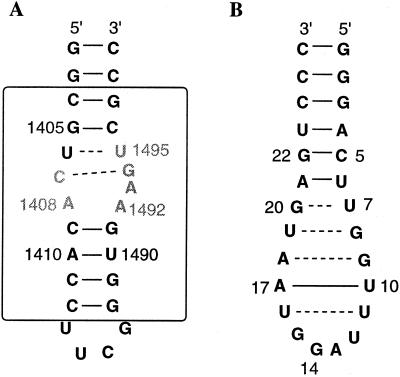
(A) Model of the A-site RNA template used to study the interactions of paromomycin. The box represents the portion of the rRNA that is homologous to the A-site. (B) RNA aptamer template used to study the interactions of tobramycin.
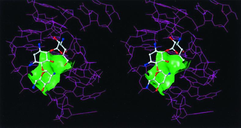
Stereo view of the complex of tobramycin bound to the RNA aptamer. The green Connolly surface represents a portion of the aminoglycoside binding site.
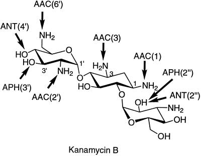
Sites of modification on kanamycin B by various aminoglycoside-modifying enzymes. The arrows point to the sites of modification by the specific enzymes, namely, acetyltransferases, phosphotransferases, and nucleotidyltransferases.
References
-
- Alper P B, Hendrix M, Sears P, Wong C-H. Probing the specificity of aminoglycoside-ribosomal RNA interactions with designed synthetic analogs. J Am Chem Soc. 1998;120:1965–1978.
-
- Azucena E, Grapsas I, Mobashery S. Properties of a bifunctional bacterial antibiotic resistance enzyme that catalyzes ATP-dependent 2"-phosphorylation and acetyl-CoA-dependent 6′-acetylation of aminoglycosides. J Am Chem Soc. 1997;119:2317–2318.
-
- Beauclerk A A, Cundliffe E. Sites of action of two ribosomal RNA methylases responsible for resistance to aminoglycosides. J Mol Biol. 1987;193:661–671. - PubMed
-
- Cate J H, Yusupov M M, Yusupova G Z, Earnest T E, Noller H F. X-ray crystal structure of 70S ribosome functional complexes. Science. 1999;285:2095–2104. - PubMed
Publication types
MeSH terms
Substances
LinkOut - more resources
Full Text Sources
Other Literature Sources
Medical
