Review
. 2000 Dec;84(6):670-3.
doi: 10.1136/heart.84.6.670.

Clinical anatomy of the aortic root

Affiliations
Review

Clinical anatomy of the aortic root

R H Anderson. Heart. 2000 Dec.
No abstract available


Figures

Figure 1:
Figure 1:
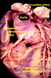
In this normal human heart, viewed in attitudinally correct orientation, the subpulmonary infundibulum has been transected, and the pulmonary valve removed, showing the central position of the aortic root within the cardiac short axis.
Figure 2:
Figure 2:
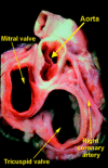
The ventricular apexes have been amputated from this ventricular mass, and the base of the heart is shown from beneath in left anterior oblique orientation. Note the central location of the aortic valve, which is overlapped by the mitral valve within the short axis of the left ventricle. The tricuspid and pulmonary valves are separated by the supraventricular crest in the roof of the right ventricle. The dotted line shows the area of fibrous continuity between the leaflets of the aortic and mitral valves.
Figure 3:
Figure 3:
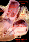
The short axis of the heart is photographed from above and from the right, producing a right posterior oblique orientation. Note that the aortic root is deeply wedged between the orifices of the mitral and tricuspid valves. Note also the origins of the coronary arteries from the sinuses of the aortic valve adjacent to the pulmonary trunk.
Figure 4:
Figure 4:
This is a close up of the aortic root, having sectioned the left ventricle along its own long axis. Note the length of the root between the basal attachments of the valvar leaflets and the sinutubular junction. Note also the thin areas of aortic wall which separate the left ventricular cavity from the pericardial space just below the level of the sinutubular junction.
Figure 5:
Figure 5:
The aortic root has been opened through a longitudinal incision across the area of aortic-mitral valvar continuity, and spread open to show the semilunar attachments of the valvar leaflets. Note the interleaflet triangles extending to the sinutubular junction, and the crescents of myocardium at the base of the two coronary aortic sinuses.
Figure 6:
Figure 6:
This section across one of the two coronary sinuses of the aortic valve shows how the hinge of the valvar leaflet is attached to the ventricular myocardium well proximal to the anatomic ventriculo-arterial junction (see fig 5 also).
Figure 7:
Figure 7:
A diagrammatic representation of the aortic root shows its considerable length. The leaflets are attached within the cylinder of the root in the form of a coronet.

References

    1. Ann Thorac Surg. 1995 Feb;59(2):419-27 - PubMed
    1. Ann Thorac Surg. 1999 Sep;68(3 Suppl):S37-43 - PubMed
    1. J Am Coll Cardiol. 1998 May;31(6):1420-5 - PubMed
    1. J Thorac Cardiovasc Surg. 1997 Jul;114(1):16-24 - PubMed

Publication types

MeSH terms