. 2000 Dec;68(12):6602-10.
doi: 10.1128/IAI.68.12.6602-6610.2000.

Dual role of signaling pathways leading to Ca(2+) and cyclic AMP elevation in host cell invasion by Trypanosoma cruzi

Affiliations

Dual role of signaling pathways leading to Ca(2+) and cyclic AMP elevation in host cell invasion by Trypanosoma cruzi

E V Caler et al. Infect Immun. 2000 Dec.

Abstract

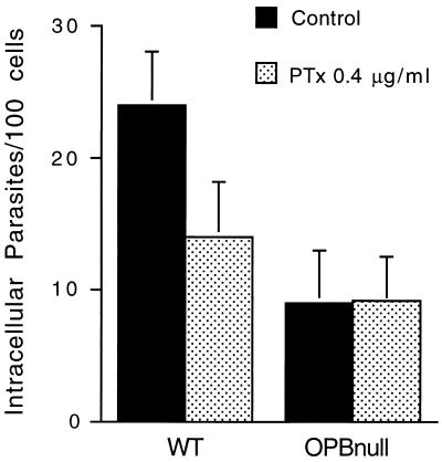
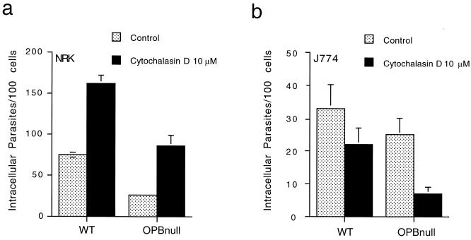
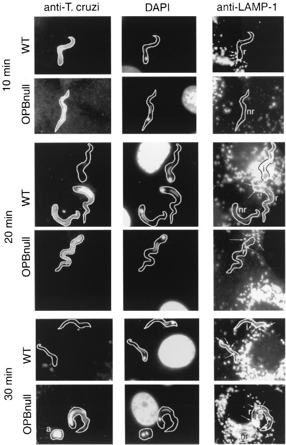
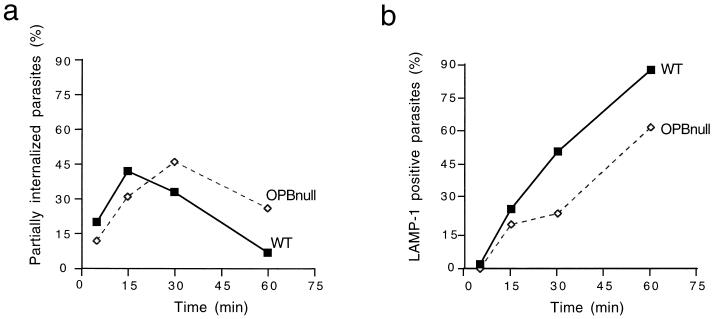
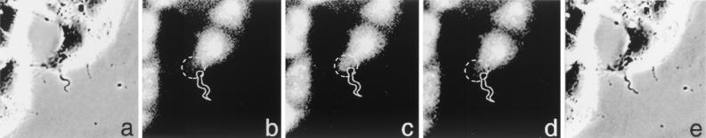
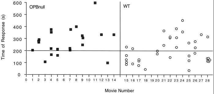
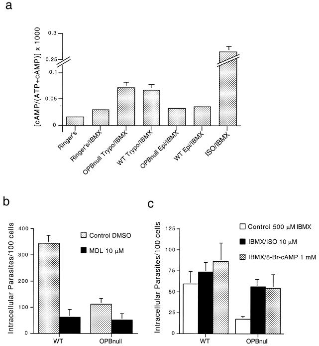
Cell invasion by the protozoan parasite Trypanosoma cruzi involves activation of host signaling pathways and the recruitment and fusion of lysosomes at the parasite entry site. A major signaling pathway regulating invasion of fibroblasts, epithelial cells, and myoblasts involves mobilization of Ca(2+) from intracellular stores and requires the activity of a T. cruzi serine peptidase, oligopeptidase B (OPB). Deletion of the OPB gene results in a marked defect in trypomastigote virulence, consistent with a greatly reduced cell invasion capacity. Here we show that uptake by macrophages, on the other hand, is largely independent of OPB expression and sensitive to inhibition of by cytochalasin D. The residual invasion capacity of OPBnull trypomastigotes in fibroblasts still involves lysosome recruitment, although in a significantly delayed fashion. Transient elevations in intracellular Ca(2+) concentrations were observed in host cells exposed to both wild-type and OPBnull trypomastigotes, but the signals triggered by the mutant parasites were less vigorous and delayed. The capacity of triggering elevation in host cell cyclic AMP (cAMP), however, was unaltered in OPBnull trypomastigotes. Modulation in cAMP levels preferentially affected the residual cell invasion capacity of OPBnull parasites, suggesting that this signaling pathway can play a dominant role in promoting cell invasion in the absence of the major OPB-dependent pathway.


Figures

FIG. 1
FIG. 1
PTx treatment of host cells does not affect the residual cell invasion capacity of OPBnull trypomastigotes. NRK fibroblasts were pretreated or not with 0.4 mg Ptx per ml overnight at 37°C. After treatment, the toxin was removed and the cells were exposed to WT or OPBnull trypomastigotes for 30 min. The data represent the average of triplicates ± the standard deviation (SD).
FIG. 2
FIG. 2
Cytochalasin D enhances invasion of NRK cells and inhibits invasion of J774 macrophages by WT and OPBnull trypomastigotes. Cells were pretreated with 10 μM cytochalasin D for 10 min at 37°C and then exposed to parasites for 1 h (a, NRK) or 30 min (b, J774). The data represent the average of triplicates ± the SD.
FIG. 3
FIG. 3
Kinetics of lysosome recruitment by WT and OPBnull trypomastigotes in NRK fibroblasts. NRK cells were exposed to 108 WT or OPBnull trypomastigotes per ml for 10, 20, or 30 min at 37°C. Immunofluorescence was performed using (i) anti-T. cruzi antibodies prior to cell permeabilization to detect extracellular parasites (left column), (ii) DAPI to detect NRK and parasite nuclei (center column) or (iii) anti-Lamp-1 monoclonal antibody after cell permeabilization to detect host cell lysosomes (right column). Lysosomes recruited to the sites of parasite attachment are indicated by arrows. i, Intracellular parasites; r, extracellular parasites recruiting lysosomes; nr, extracellular parasites not recruiting lysosomes.
FIG. 4
FIG. 4
OPBnull trypomastigotes recruit lysosomes and invade cells in a delayed pattern. NRK cells were exposed to WT or OPBnull trypomastigotes for the indicated periods of time. Parasites attached to the cells and partially internalized were stained with an anti-T. cruzi antibody; recruitment of lysosomes was visualized by staining with an anti-Lamp-1 monoclonal antibody after cell permeabilization, and the total number of parasites was determined by nuclear staining with DAPI. (a) Percent parasites recruiting lysosomes = (number of anti-T. cruzi-Lamp-1 double-positive parasites/total parasites) × 100. (b) Percent internalized parasites = (Lamp-1-positive parasites/total parasites) × 100.
FIG. 5
FIG. 5
Localized Ca2+ signaling response generated by T. cruzi trypomastigotes in NRK cells. (a) Phase-contrast image of a stably attached trypomastigote at the beginning of the time lapse recording. (b, c, and d) Fluorescence images of fluo-3-loaded NRK cells at 39, 41, and 43 s, respectively, after initiation of the time-lapse recording, showing a localized transient [Ca2+]i elevation at the site of parasite attachment. (e) Phase-contrast image obtained after finalization of the time-lapse recording. Dashed circles indicate the region of the cell were a localized [Ca2+]i elevation occurred as a consequence of parasite attachment.
FIG. 6
FIG. 6
OPBnull trypomastigotes trigger delayed Ca2+ signaling in NRK cells. WT (■) or OPBnull (□) trypomastigotes were added to NRK cells preloaded with the Ca2+-sensitive dye fluo-3. Time-lapse images were acquired, and the time frame in which each individual [Ca2+]i elevation occurred was determined. The plot shows the total number of responsive cells in each individual movie for a recording period of 600 s.
FIG. 7
FIG. 7
Residual cell invasion by OPBnull trypomastigotes is modulated by cAMP. (a) Intracellular levels of cAMP were measured in IBMX-pretreated NRK cells after exposure to WT and OPBnull trypomastigotes and epimastigotes or exposure to isoproterenol. Infection of NRK cells by OPBnull trypomastigotes was quantitated after pretreatment of the host cells with different cAMP modulators: b, 10 μM MDL-12,330A for 30 min at 37°C; and c, 10 μM isoproterenol or 1 mM 8-Br-cAMP for 30 min at 37°C. The data represent the average of triplicate determinations ± the SD.

References

    1. Sibley L D, Andrews N W. Cell invasion by un-palatable parasites. Traffic. 2000;1:100–106. - PubMed
    1. Andrews N W. Lysosome recruitment during host cell invasion by Trypanosoma cruzi. Trends Cell Biol. 1995;5:133–137. - PubMed
    1. Andrews N W, Hong K S, Robbins E S, Nussenzweig V. Stage-specific surface antigens expressed during the morphogenesis of vertebrate forms of Trypanosoma cruzi. Exp Parasitol. 1987;64:474–484. - PubMed
    1. Burleigh B A, Andrews N W. A 120-kDa alkaline peptidase from Trypanosoma cruzi is involved in the generation of a novel Ca2+-signaling factor for mammalian cells. J Biol Chem. 1995;270:5172–5180. - PubMed
    1. Burleigh B A, Caler E V, Webster P, Andrews N W. A cytosolic serine endopeptidase from Trypanosoma cruzi is required for the generation of Ca2+ signaling in mammalian cells. J Cell Biol. 1997;136:609–620. - PMC - PubMed

Publication types

LinkOut - more resources