. 2000 Dec;68(12):6903-11.
doi: 10.1128/IAI.68.12.6903-6911.2000.

Cellular internalization of cytolethal distending toxin from Haemophilus ducreyi

Affiliations

Cellular internalization of cytolethal distending toxin from Haemophilus ducreyi

X Cortes-Bratti et al. Infect Immun. 2000 Dec.

Abstract

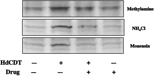
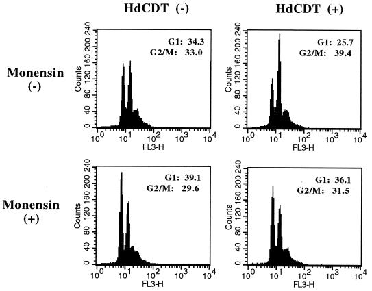
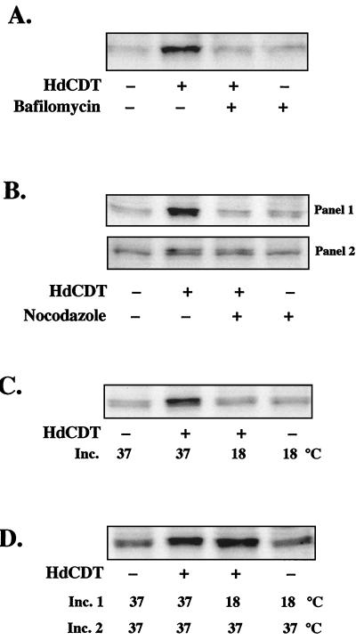
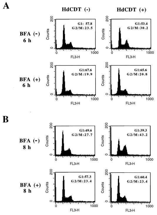
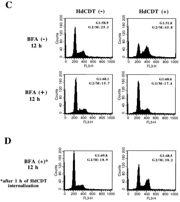
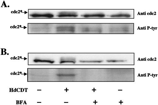
The chancroid bacterium Haemophilus ducreyi produces a toxin (HdCDT) which is a member of the recently discovered family of cytolethal distending toxins (CDTs). These protein toxins prevent the cyclin-dependent kinase cdc2 from being activated, thus blocking the transition of cells from the G(2) phase into mitosis, with the consequent arrest of intoxicated cells in G(2). It is not known whether these toxins act by signaling from the cell surface or intracellularly only. Here we report that HdCDT has to undergo at least internalization before being able to act. Cellular intoxication was inhibited (i) by removal of clathrin coats via K(+) depletion, (ii) by treatment with drugs that inhibit receptor clustering into coated pits, and (iii) in cells genetically manipulated to fail in clathrin-dependent endocytosis. Intoxication was also completely inhibited in cells treated with bafilomycin A1 or nocodazole and in cells incubated at 18 degrees C, i.e., under conditions known to block the fusion of early endosomes with downstream compartments. Moreover, disruption of the Golgi complex by treatment with brefeldin A or ilimaquinone blocked intoxication. In conclusion, our data indicate that HdCDT enters cells via clathrin-coated pits and has to be transported via the Golgi complex in order to intoxicate cells. This is the first member of the family of CDTs for which cellular internalization and some details of the pathway have been demonstrated.


Figures

FIG. 1
FIG. 1
Effects on HdCDT-induced intoxication of drugs which inhibit endosomal acidification. Cells were preincubated with methylamine (10 mM), NH4Cl (20 mM), or monensin (10 μM), treated with the toxin for 15 min, and postincubated for 12 h in the presence of the respective drug. Tyrosine phosphorylation of cdc2 in the treated cells was determined by Western blotting as described in Materials and Methods. Blots are representative of two different experiments with each drug.
FIG. 2
FIG. 2
Effect of monensin on HdCDT-induced intoxication. Flow cytometry was used to analyze control cells [HdCDT(−)] and toxin-treated cells [HdCDT (+)] in the presence (+) or absence (−) of monensin (10 μM). Samples were prepared 12 h after toxin treatment, and DNA was stained with propidium iodide. The G1 and G2/M regions were marked, and the percentages of cells in these phases are shown. One representative experiment of two is shown. FL3-H, relative fluorescence.
FIG. 3
FIG. 3
Effect of K+ depletion and chlorpromazine treatment on tyrosine phosphorylation of cdc2 in HdCDT-treated cells. Cells were depleted of K+ as described in Materials and Methods and treated with HdCDT; samples were prepared 24 h after toxin exposure. For chlorpromazine treatment, cells were preincubated with chlorpromazine (25 μg/ml, 1 h), treated with HdCDT, and postincubated for 8 h with 10 μg of the drug per ml. Intoxication with DT and measurement of DT activity (inset) were performed as described in Materials and Methods. Error bars indicate standard deviations.
FIG. 4
FIG. 4
Effect of HdCDT on HeLadynK44A cells. Tyrosine phosphorylation of cdc2 after HdCDT treatment was measured in cells expressing only endogenous dynamin (tetracycline +) and in cells overexpressing dominant-negative dynamin (tetracycline −). To obtain overexpression, cells were grown in the absence of tetracycline for 2 days before the toxin treatment. Samples were taken 24 h after toxin exposure. Blots are representative of four different experiments. P-Tyr, phosphotyrosine.
FIG. 5
FIG. 5
Effects of BafA1 (50 μM), nocodazole (30 μM), and 18°C incubation on tyrosine phosphorylation of cdc2 in HdCDT-treated cells. (A) Cells were exposed to BafA1 before and after toxin treatment. The postincubation time was 24 h. (B) Cells were exposed to nocodazole before and directly after toxin treatment (panel 1) or received nocodazole 60 min after toxin treatment (panel 2). The postincubation time was 8 h. (C and D) Cells were cultivated in eight individual petri dishes; four were kept as controls, and four were exposed to the toxin at the same time. (C) One pair of plates (HdCDT − and HdCDT +) was incubated (Inc.) at 37°C and the other was incubated at 18°C. Samples were prepared 12 h after toxin treatment. (D) One pair of plates was incubated at 37°C for 24 h. The other was incubated at 18°C for the first 12 h (Inc. 1) and then transferred to 37°C for another 12 h (Inc. 2). Blots are representative of three different experiments with each treatment.
FIG. 6
FIG. 6
Effect of BFA on HdCDT-induced intoxication. Flow cytometry was used to analyze control [HdCDT (−)] and toxin-treated [HdCDT (+)] cells in the presence (+) or absence (−) of BFA. Cells were pretreated with BFA (2.5 μg/ml) for 45 min, exposed to the toxin, and postincubated in normal medium with BFA. Samples were prepared 6 h (A), 8 h (B), and 12 h (C) after toxin treatment. (D) Sample from cells treated with toxin, postincubated for 45 min at 37°C in fresh medium to allow internalization of the toxin, and exposed for 12 h to BFA. Percentages of cells in G1 and G2/M are shown. One representative experiment of three is shown. FL3-H, relative fluorescence.
FIG. 6
FIG. 6
Effect of BFA on HdCDT-induced intoxication. Flow cytometry was used to analyze control [HdCDT (−)] and toxin-treated [HdCDT (+)] cells in the presence (+) or absence (−) of BFA. Cells were pretreated with BFA (2.5 μg/ml) for 45 min, exposed to the toxin, and postincubated in normal medium with BFA. Samples were prepared 6 h (A), 8 h (B), and 12 h (C) after toxin treatment. (D) Sample from cells treated with toxin, postincubated for 45 min at 37°C in fresh medium to allow internalization of the toxin, and exposed for 12 h to BFA. Percentages of cells in G1 and G2/M are shown. One representative experiment of three is shown. FL3-H, relative fluorescence.
FIG. 7
FIG. 7
Effect of BFA on the tyrosine phosphorylation of cdc2 in HdCDT-treated cells 8 h (A) and 12 h (B) after toxin treatment. Blots are representative of three different experiments. P-tyr, phosphotyrosine.
FIG. 8
FIG. 8
Effect of ilimaquinone (Ilimaq.) on HdCDT-induced intoxication. Shown are Flow cytometric analysis (A) and tyrosine phosphorylation of cdc2 (B) in control [HdCDT (−)] and toxin-treated [HdCDT (+)] cells with (+) or without (−) ilimaquinone. One representative experiment of two is shown.

References

    1. Bayer N, Schober D, Prchla E, Murphy R F, Blaas D, Fuchs R. Effect of bafilomycin A1 and nocodazole on endocytic transport in HeLa cells: implications for viral uncoating and infection. J Virol. 1998;72:9645–9655. - PMC - PubMed
    1. Chardin P, McCormick F. Brefeldin A: the advantage of being uncompetitive. Cell. 1999;97:153–155. - PubMed
    1. Cieplak W, Jr, Messer R J, Konkel M E, Grant C C R. Role of potential endoplasmic reticulum retention sequence (RDEL) and the Golgi complex in the cytotonic activity of Escherichia coli heat-labile enterotoxin. Mol Microbiol. 1995;16:789–800. - PubMed
    1. Comayras C, Tasca C, Peres S Y, Ducommun B, Oswald E, De Rycke J. Escherichia coli cytolethal distending toxin blocks the HeLa cell cycle at the G2/M transition by preventing cdc2 protein kinase dephosphorylation and activation. Infect Immun. 1997;65:5088–5095. - PMC - PubMed
    1. Cope L D, Lumbley S, Latimer J L, Klesney-Tait J, Stevens M K, Johnson L S, Purvén M, Munson R S, Jr, Lagergård T, Radolf J D, Hansen E J. A diffusible cytotoxin of Haemophilus ducreyi. Proc Natl Acad Sci USA. 1997;94:4056–4061. - PMC - PubMed

Publication types

LinkOut - more resources