. 2000 Dec;68(12):6939-45.
doi: 10.1128/IAI.68.12.6939-6945.2000.

Phagocytic uptake of Encephalitozoon cuniculi by nonprofessional phagocytes

Affiliations

Phagocytic uptake of Encephalitozoon cuniculi by nonprofessional phagocytes

S Couzinet et al. Infect Immun. 2000 Dec.

Abstract

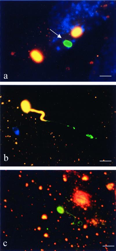
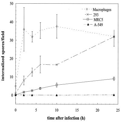
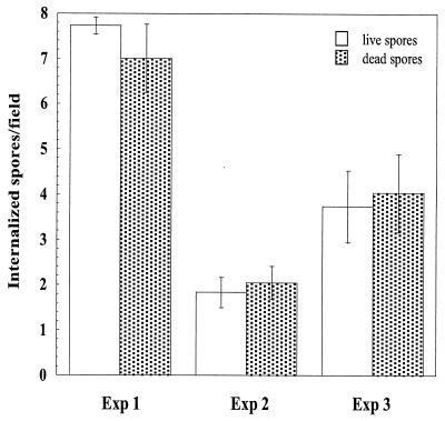
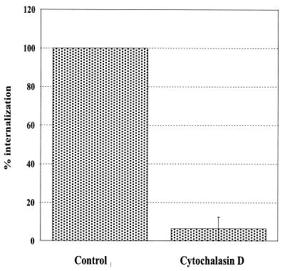
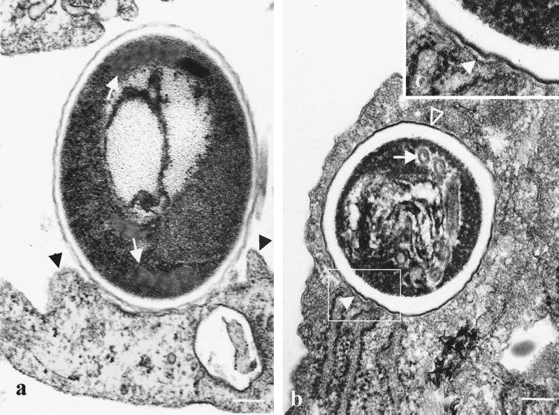
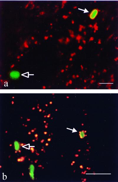
Encephalitozoon cuniculi is an obligate intracellular, spore-forming parasite belonging to the microsporidia that can cause disseminated infection in immunocompromised persons. E. cuniculi spores infect host cells by germination, i.e., by explosively everting the polar filament, through which the spore contents (sporoplasms) are subsequently injected into the cytoplasm. In addition, we observed intracellular, nongerminated spores in various nonprofessional phagocytes. In MRC5 cells, the number of internalized spores was approximately 10-fold higher than the number of injected sporoplasms. Compared to the rate of uptake by human monocyte-derived macrophages, internalization rates by A549 cells, MRC5 cells, and 293 cells were 0.6, 4.4, and 22.2%, respectively. The mechanism of uptake was studied in MRC5 cells. Killed spores were internalized at the same rate as live spores, indicating that nongerminated parasites do not actively participate in cell entry. Cytochalasin D inhibited uptake of spores by 95%, demonstrating an actin-dependent process. By electron and epifluorescence microscopy, intracellular spores were found in a tightly fitting membrane-bound compartment. The vacuole containing the spores was positive for the lysosomal membrane protein LAMP-1 and colocalized with the late endosomal-lysosomal content marker rhodamine dextran. Our results show that, in addition to the unique way in which microsporidia infect cells, E. cuniculi spores enter nonprofessional phagocytes by phagocytosis and traffic into a late endosomal-lysosomal compartment.


Figures

FIG. 1
FIG. 1
Differential immunofluorescence staining of MRC5 cells coincubated with E. cuniculi. The first labeling, using an anti-E. cuniculi antibody and a Cy3-conjugated secondary antibody (orange fluorescence), was performed without permeabilizing the host cells in order to label only extracellular spores. Host cells were then permeabilized with saponin followed by labeling with the same anti-E. cuniculi antibody visualized by a FITC-conjugated secondary antibody (green fluorescence). Extracellular microsporidia (spore walls and polar filaments) appear orange and intracellular ones appear green. Nuclei of MRC5 cells are stained blue (revealed by DAPI). (a) Intracellular, nongerminated spore (arrow). (b) Germinated extracellular spore inserting terminal portion of polar filament into host cell. (c) Germinated intracellular spore. Spore wall and entire polar filament are intracellular. Bar = 3 μm.
FIG. 2
FIG. 2
Kinetics of the internalization of nongerminated E. cuniculi spores by various NPP cell lines and human MDM. Two million E. cuniculi spores were added to each well of confluent cells, and the monolayers were fixed after 0, 2, 4, 6, 10, and 24 h. Intracellular spores were identified using differential immunofluorescence staining. To account for the size difference between the cells studied, internalization rates are given as the number of intracellular spores per high-power field of view (magnification, ×1,000). In confluent monolayers every field covers approximately 11.5 MRC5 cells, 50 293 cells, 60 A549 cells, and 30 macrophages. Data shown are means ± SD for three independent experiments, except for time points of >6 h for 293 cells, where the results of a single experiment are given (monolayers of 293 cells tended to detach from the coverslip after >6 h). Differences between cell lines were significant at a P value of <0.0001 (two-way analysis of variance).
FIG. 3
FIG. 3
Internalization of live and dead E. cuniculi spores by MRC5 cells. Two million either live or formaldehyde-fixed spores were added to each well of confluent cells and coincubated for 2 h. Intracellular spores were identified using differential immunofluorescence staining. The rate of uptake of spores differed markedly between experiments. However, there was no statistically significant difference in the rate of uptake between live and formaldehyde-fixed spores (P ≫ 0.05 by Student's t test). Exp, experiment.
FIG. 4
FIG. 4
The role of actin polymerization in the internalization of E. cuniculi spores. The uptake of nongerminated spores by MRC5 cells in the presence of 10 μg of cytochalasin D per ml is compared to that for concurrently performed control assays in the absence of cytochalasin D. Data are expressed as the means ± SD of three experiments (P = 0.03 by paired Student's t test).
FIG. 5
FIG. 5
Electron micrographs of MRC5 cells coincubated with E. cuniculi spores for 2 h. (a) Tight association of the bound spore with the cell membrane and membrane protrusions resembling pseudopods extended along the spore wall (black arrowheads) are observed. Cross sections of coiled polar filament can also be seen (white arrows). (b) Characteristic electron-dense outer (open arrowhead) and electron-lucent inner layers of the wall of a mature E. cuniculi spore, as well as various sections of coils of the polar tube (white arrow), are shown. (c) Detail of the enclosed area from panel b showing a membrane tightly apposed to the contour of the internalized spore (closed arrowhead). Bar = 0.2 μm.
FIG. 6
FIG. 6
Colocalization of late endosomal-lysosomal markers with internalized E. cuniculi spores. (a) Lysosomal membrane protein LAMP-1 revealed by immunofluorescence. MRC5 cells were coincubated with spores for 2 h, fixed, and simultaneously labeled with a rabbit anti-E. cuniculi antibody and a mouse anti-LAMP-1 monoclonal antibody visualized by a FITC-conjugated and a Cy3-conjugated secondary antibody, respectively. The internalized spore (closed arrow) is surrounded by a ring of orange fluorescence (LAMP-1), indicating fusion of the vesicle containing the spore with late endosomes and lysosomes. LAMP-1 does not colocalize with the extracellular spore (open arrow). (b) RD localization by fluorescence. Cells were incubated with RD for 24 h, washed, and kept in RD-free medium for 2 h to allow passage of the marker to the late endosomal-lysosomal compartment. The RD-labeled cells were then coincubated with spores for 30 min, fixed, and labeled with an anti-E. cuniculi antibody visualized by a FITC-conjugated secondary antibody. The phagocytosed spore (closed arrow) surrounded by red fluorescent marker indicates phagosome-lysosome fusion. Extracellular spores are not associated with RD (open arrow). Bar = 3 μm.

References

    1. Aderem A, Underhill D M. Mechanisms of phagocytosis in macrophages. Annu Rev Immunol. 1999;7:593–623. - PubMed
    1. Böyum A. Isolation of mononuclear cells and granulocytes from human blood. Isolation of monuclear cells by one centrifugation, and of granulocytes by combining centrifugation and sedimentation at 1 g. Scand J Clin Lab Investig Suppl. 1968;97:77–89. - PubMed
    1. Canning E, Lom J, Dykova I. The microsporidia of vertebrates. New York, N.Y: Academic Press, Inc.; 1986.
    1. Canning E U. Microsporidia. In: Kreier J P, Vaker J R, editors. Parasitic protozoa. 2nd ed. Vol. 6. New York, N.Y: Academic Press; 1993. pp. 299–385.
    1. Chi E, Mehl T, Nunn D, Lory S. Interaction of Pseudomonas aeruginosa with A549 pneumocyte cells. Infect Immun. 1991;59:822–828. - PMC - PubMed

Publication types

LinkOut - more resources