

Immunity to murine Chlamydia trachomatis genital tract reinfection involves B cells and CD4(+) T cells but not CD8(+) T cells
- PMID: 11083822
- PMCID: PMC97807
- DOI: 10.1128/IAI.68.12.6979-6987.2000
Immunity to murine Chlamydia trachomatis genital tract reinfection involves B cells and CD4(+) T cells but not CD8(+) T cells
Abstract
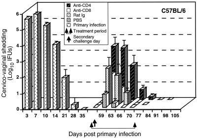
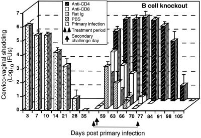
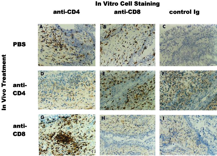
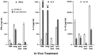
CD4(+) T-helper type 1 (Th1) responses are essential for the resolution of a primary Chlamydia trachomatis genital tract infection; however, elements of the immune response that function in resistance to reinfection are poorly understood. Defining the mechanisms of immune resistance to reinfection is important because the elements of protective adaptive immunity are distinguished by immunological memory and high-affinity antigen recognition, both of which are crucial to the development of efficacious vaccines. Using in vivo antibody depletion of CD4(+) and CD8(+) T cells prior to secondary intravaginal challenge, we identified lymphocyte populations that functioned in resistance to secondary chlamydial infection of the genital tract. Depletion of either CD4(+) or CD8(+) T cells in immune wild-type C57BL/6 mice had a limited effect on resistance to reinfection. However, depletion of CD4(+) T cells, but not CD8(+) T cells, in immune B-cell-deficient mice profoundly altered the course of secondary infection. CD4-depleted B-cell-deficient mice were unable to resolve a secondary infection, shed high levels of infectious chlamydiae, and did not resolve the infection until 3 to 4 weeks following the discontinuation of anti-CD4 treatment. These findings substantiated a predominant role for CD4(+) T cells in host resistance to chlamydial reinfection of the female genital tract and demonstrated that CD8(+) T cells are unnecessary for adaptive immune resistance. More importantly, however, this study establishes a previously unrecognized but very significant role for B cells in resistance to chlamydial reinfection and suggests that B cells and CD4(+) T cells may function synergistically in providing immunity in this model of chlamydial infection. Whether CD4(+) T cells and B cells function independently or dependently is unknown, but definition of those mechanisms is fundamental to understanding optimum protective immunity and to the development of highly efficacious immunotherapies against chlamydial urogenital infections.
Figures

Similar articles
-
Resolution of secondary Chlamydia trachomatis genital tract infection in immune mice with depletion of both CD4+ and CD8+ T cells.Infect Immun. 2001 Apr;69(4):2643-9. doi: 10.1128/IAI.69.4.2643-2649.2001. Infect Immun. 2001. PMID: 11254630 Free PMC article.
-
A predominant role for antibody in acquired immunity to chlamydial genital tract reinfection.J Immunol. 2005 Dec 1;175(11):7536-42. doi: 10.4049/jimmunol.175.11.7536. J Immunol. 2005. PMID: 16301662 Free PMC article.
-
Immunological memory in B-cell-deficient mice conveys long-lasting protection against genital tract infection with Chlamydia trachomatis by rapid recruitment of T cells.Immunology. 2001 Feb;102(2):199-208. doi: 10.1046/j.1365-2567.2001.01167.x. Immunology. 2001. PMID: 11260325 Free PMC article.
-
Vaccines for Chlamydia infections of the female genital tract.Future Microbiol. 2008 Feb;3(1):67-77. doi: 10.2217/17460913.3.1.67. Future Microbiol. 2008. PMID: 18230035 Review.
-
T cell responses to Chlamydia.Pathog Dis. 2021 Mar 31;79(4):ftab014. doi: 10.1093/femspd/ftab014. Pathog Dis. 2021. PMID: 33693620 Free PMC article. Review.
Cited by
-
Animal models for studying female genital tract infection with Chlamydia trachomatis.Infect Immun. 2013 Sep;81(9):3060-7. doi: 10.1128/IAI.00357-13. Epub 2013 Jul 8. Infect Immun. 2013. PMID: 23836817 Free PMC article. Review.
-
Dynamics of NKT-Cell Responses to Chlamydial Infection.Front Immunol. 2015 May 15;6:233. doi: 10.3389/fimmu.2015.00233. eCollection 2015. Front Immunol. 2015. PMID: 26029217 Free PMC article. Review.
-
Human Fallopian Tube Epithelial Cell Culture Model To Study Host Responses to Chlamydia trachomatis Infection.Infect Immun. 2020 Aug 19;88(9):e00105-20. doi: 10.1128/IAI.00105-20. Print 2020 Aug 19. Infect Immun. 2020. PMID: 32601108 Free PMC article.
-
Chlamydia and Its Many Ways of Escaping the Host Immune System.J Pathog. 2019 Aug 6;2019:8604958. doi: 10.1155/2019/8604958. eCollection 2019. J Pathog. 2019. PMID: 31467721 Free PMC article. Review.
-
Immunization with the Chlamydia trachomatis mouse pneumonitis major outer membrane protein can elicit a protective immune response against a genital challenge.Infect Immun. 2001 Oct;69(10):6240-7. doi: 10.1128/IAI.69.10.6240-6247.2001. Infect Immun. 2001. PMID: 11553566 Free PMC article.
References
Publication types
MeSH terms
Grants and funding
LinkOut - more resources
Full Text Sources
Other Literature Sources
Medical
Research Materials
