Identification of discrete domains within gonococcal transferrin-binding protein A that are necessary for ligand binding and iron uptake functions
- PMID: 11083823
- PMCID: PMC97808
- DOI: 10.1128/IAI.68.12.6988-6996.2000
Identification of discrete domains within gonococcal transferrin-binding protein A that are necessary for ligand binding and iron uptake functions
Abstract
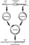
The availability of free iron in vivo is strictly limited, in part by the iron-binding protein transferrin. The pathogenic Neisseria spp. can sequester iron from this protein, dependent upon two iron-repressible, transferrin-binding proteins (TbpA and TbpB). TbpA is a TonB-dependent, integral, outer membrane protein that may form a beta-barrel exposing multiple surface loops, some of which are likely to contain ligand-binding motifs. In this study we propose a topological model of gonococcal TbpA and then test some of the hypotheses set forth by the model by individually deleting three putative loops (designated loops 4, 5, and 8). Each mutant TbpA could be expressed without toxicity and was surface exposed as assessed by immunoblotting, transferrin binding, and protease accessibility. Deletion of loop 4 or loop 5 abolished transferrin binding to whole cells in solid- and liquid-phase assays, while deletion of loop 8 decreased the affinity of the receptor for transferrin without affecting the copy number. Strains expressing any of the three mutated TbpAs were incapable of growth on transferrin as a sole iron source. These data implicate putative loops 4 and 5 as critical determinants for receptor function and transferrin-iron uptake by gonococcal TbpA. The phenotype of the DeltaL8TbpA mutant suggests that high-affinity ligand interaction is required for transferrin-iron internalization.
Figures


Similar articles
-
Energy-dependent changes in the gonococcal transferrin receptor.Mol Microbiol. 1997 Oct;26(1):25-35. doi: 10.1046/j.1365-2958.1997.5381914.x. Mol Microbiol. 1997. PMID: 9383187
-
Identification of TbpA residues required for transferrin-iron utilization by Neisseria gonorrhoeae.Infect Immun. 2008 May;76(5):1960-9. doi: 10.1128/IAI.00020-08. Epub 2008 Mar 17. Infect Immun. 2008. PMID: 18347046 Free PMC article.
-
Demonstration and characterization of a specific interaction between gonococcal transferrin binding protein A and TonB.J Bacteriol. 2002 Nov;184(22):6138-45. doi: 10.1128/JB.184.22.6138-6145.2002. J Bacteriol. 2002. PMID: 12399483 Free PMC article.
-
Identification of receptor-mediated transferrin-iron uptake mechanism in Neisseria gonorrhoeae.Methods Enzymol. 1994;235:356-63. doi: 10.1016/0076-6879(94)35154-6. Methods Enzymol. 1994. PMID: 8057908 Review. No abstract available.
-
Transferrin-iron uptake by Gram-negative bacteria.Front Biosci. 2003 May 1;8:d836-47. doi: 10.2741/1076. Front Biosci. 2003. PMID: 12700102 Review.
Cited by
-
Determination of surface-exposed, functional domains of gonococcal transferrin-binding protein A.Infect Immun. 2004 Mar;72(3):1775-85. doi: 10.1128/IAI.72.3.1775-1785.2004. Infect Immun. 2004. PMID: 14977987 Free PMC article.
-
An immunogenic, surface-exposed domain of Haemophilus ducreyi outer membrane protein HgbA is involved in hemoglobin binding.Infect Immun. 2009 Jul;77(7):3065-74. doi: 10.1128/IAI.00034-09. Epub 2009 May 18. Infect Immun. 2009. PMID: 19451245 Free PMC article.
-
Investigating the importance of selected surface-exposed loops in HpuB for hemoglobin binding and utilization by Neisseria gonorrhoeae.Infect Immun. 2024 Jul 11;92(7):e0021124. doi: 10.1128/iai.00211-24. Epub 2024 Jun 12. Infect Immun. 2024. PMID: 38864605 Free PMC article.
-
A structural comparison of human serum transferrin and human lactoferrin.Biometals. 2007 Jun;20(3-4):249-62. doi: 10.1007/s10534-006-9062-7. Epub 2007 Jan 11. Biometals. 2007. PMID: 17216400 Free PMC article. Review.
-
The transferrin-iron import system from pathogenic Neisseria species.Mol Microbiol. 2012 Oct;86(2):246-57. doi: 10.1111/mmi.12002. Epub 2012 Sep 7. Mol Microbiol. 2012. PMID: 22957710 Free PMC article. Review.
References
-
- Biswas G D, Anderson J E, Sparling P F. Cloning and functional characterization of Neisseria gonorrhoeae tonB, exbB, and exbD genes. Mol Microbiol. 1997;24:169–179. - PubMed
Publication types
MeSH terms
Substances
Grants and funding
LinkOut - more resources
Full Text Sources
Other Literature Sources
Medical
Research Materials
Miscellaneous

