. 2000 Dec;68(12):6997-7002.
doi: 10.1128/IAI.68.12.6997-7002.2000.

Identification of mycobacterial surface proteins released into subcellular compartments of infected macrophages

Affiliations

Identification of mycobacterial surface proteins released into subcellular compartments of infected macrophages

W L Beatty et al. Infect Immun. 2000 Dec.

Abstract

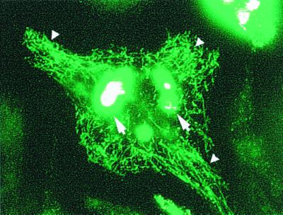
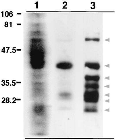
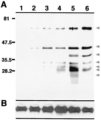
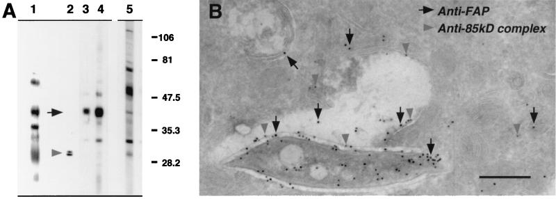
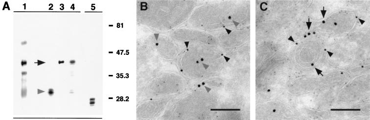
Considerable effort has focused on the identification of proteins secreted from Mycobacterium spp. that contribute to the development of protective immunity. Little is known, however, about the release of mycobacterial proteins from the bacterial phagosome and the potential role of these molecules in chronically infected macrophages. In the present study, the release of mycobacterial surface proteins from the bacterial phagosome into subcellular compartments of infected macrophages was analyzed. Mycobacterium bovis BCG was surface labeled with fluorescein-tagged succinimidyl ester, an amine-reactive probe. The fluorescein tag was then used as a marker for the release of bacterial proteins in infected macrophages. Fractionation studies revealed bacterial proteins within subcellular compartments distinct from mycobacteria and mycobacterial phagosomes. To identify these proteins, subcellular fractions free of bacteria were probed with mycobacterium-specific antibodies. The fibronectin attachment protein and proteins of the antigen 85-kDa complex were identified among the mycobacterial proteins released from the bacterial phagosome.


Figures

FIG. 1
FIG. 1
Release of labeled mycobacterial proteins from the phagosome in infected macrophages. Live BMMφ infected for 24 h with fluorescein succinimidyl ester-labeled BCG were analyzed by fluorescence microscopy. Labeled bacterial proteins were released from the mycobacterial phagosome into subcellular compartments of the infected macrophage (small arrowheads). The labeled bacteria are intensely fluorescent and are indicated by the large arrows.
FIG. 2
FIG. 2
Labeled mycobacterial proteins are present in subcellular compartments of infected macrophages. BMMφ were infected with fluorescein succinimidyl ester-labeled M. bovis BCG for 24 h. The infected cell lysates were then subjected to fractionation on sucrose gradients to isolate a macrophage subcellular membrane fraction free of bacteria. The bacterium-free subcellular compartments were then analyzed for the presence of released fluorescein-labeled bacterial proteins by probing with HRP-conjugated rabbit anti-fluorescein antibody. Lane 1, fluorescein succinimidyl ester-labeled BCG; lane 2, a bacterium-free subcellular fraction isolated from fluorescein succinimidyl ester-labeled mycobacterium-infected macrophages; lane 3, mouse anti-fluorescein immunoprecipitation of a bacterium-free macrophage subcellular fraction. Arrowheads indicate bacterial proteins immunoprecipitated from macrophage subcellular organelles.
FIG. 3
FIG. 3
Temporal release of bacterial proteins from the mycobacterial phagosome. Bacterium-free subcellular organelles isolated from uninfected macrophages (lane 1) and from macrophages infected with fluorescein succinimidyl ester-labeled BCG for 30 min (lane 2), 1 h (lane 3), 4) 2 h (lane 4), 4 h (lane 5), and 12 h (lane 6) were immunoprecipitated with a mouse anti-fluorescein antibody. The resulting material was analyzed by probing a Western blot with a rabbit anti-fluorescein antibody (A) or anti-Lamp 1 (B). Lamp 1 served as an internal control for the total amount of protein present in each sample. Arrowheads indicate bacterial proteins immunoprecipitated from macrophage subcellular organelles.
FIG. 4
FIG. 4
Identification of mycobacterial proteins released from the phagosome. (A) Bacterium-free subcellular organelles of macrophages infected for 24 h with fluorescein succinimidyl ester-labeled BCG were immunoprecipitated with a mouse anti-fluorescein antibody. The resulting material was analyzed by Western blot probing with rabbit antibodies specific to fluorescein (lane 1), the antigen 85-kDa complex of mycobacteria (lane 2), mycobacterial FAP (lane 3), and M. tuberculosis H37Rv CFP (lane 4). (Lane 5) BCG was probed with anti-H37Rv CFP to illustrate the mycobacterial protein profile recognized by this antibody. (B) Cryosections of BMMφ infected for 24 h with BCG and probed with rat anti-FAP (anti-rat 18-nm colloidal gold) (solid arrows) and rabbit anti-antigen 85-kDa complex (anti-rabbit 12-nm colloidal gold) (shaded arrowheads) revealed the release of these proteins into the bacterial phagosome and compartments of the host macrophage. Bar, 0.5 μm.
FIG. 5
FIG. 5
Mycobacterial proteins traffic to late endocytic compartments. (A) Subcellular organelles of macrophages infected for 24 h with fluorescein succinimidyl ester-labeled BCG were further fractionated by DGE to isolate late endosomal/lysosomal compartments free of bacteria. Following immunoprecipitation with mouse anti-fluorescein antibody, the resulting material was analyzed by Western blot probing with rabbit antibodies specific to fluorescein (lane 1), the antigen 85-kDa complex of mycobacteria (lane 2), mycobacterial FAP (lane 3), and M. tuberculosis H37Rv CFP (lane 4). (Lane 5) The late endosomal/lysosomal fraction was probed with an antibody recognizing the β subunit of MHC class II (KL295). (B and C) Cryosections of DGE-isolated late endosome/lysosome compartments from BMMφ infected for 24 h with BCG were probed with rabbit anti-antigen 85-kDa complex (anti-rabbit 18-nm colloidal gold) (shaded arrowheads) (B) and rabbit anti-FAP (anti-rabbit 18-nm colloidal gold) (solid arrows) (C). These compartments were also probed with mouse anti-MHC class II (KL295) (anti-mouse 12-nm colloidal gold) (solid arrowheads) and an antibody to the lysosomal marker Lamp 1 (rat anti-Lamp 1; anti-rat 6-nm colloidal gold) as shown in both panels B and C. Bar, 0.1 μm.

References

    1. Abou-Zeid C, Smith I, Grange J M, Ratliff T L, Steele J, Rook G A. The secreted antigens of Mycobacterium tuberculosisand their relationship to those recognized by the available antibodies. J Gen Microbiol. 1988;134:531–538. - PubMed
    1. Armstrong J A, Hart P D. Response of cultured macrophages to Mycobacterium tuberculosis, with observations on fusion of lysosomes with phagosomes. J Exp Med. 1971;134:713–740. - PMC - PubMed
    1. Beatty W L, Rhoades E R, Ullrich H-J, Chatterjee D, Heuser J E, Russell D G. Trafficking and release of mycobacterial lipids from infected macrophages. Traffic. 2000;1:235–247. - PubMed
    1. Clemens D L, Horwitz M A. Characterization of the Mycobacterium tuberculosisphagosome and evidence that phagosomal maturation is inhibited. J Exp Med. 1995;181:257–270. - PMC - PubMed
    1. Crowle A J, Dahl R, Ross E, May M H. Evidence that vesicles containing living, virulent Mycobacterium tuberculosis or Mycobacterium aviumin cultured human macrophages are not acidic. Infect Immun. 1991;59:1823–1831. - PMC - PubMed

Publication types

Substances

LinkOut - more resources