

Differential alteration in intestinal epithelial cell expression of toll-like receptor 3 (TLR3) and TLR4 in inflammatory bowel disease
- PMID: 11083826
- PMCID: PMC97811
- DOI: 10.1128/IAI.68.12.7010-7017.2000
Differential alteration in intestinal epithelial cell expression of toll-like receptor 3 (TLR3) and TLR4 in inflammatory bowel disease
Abstract
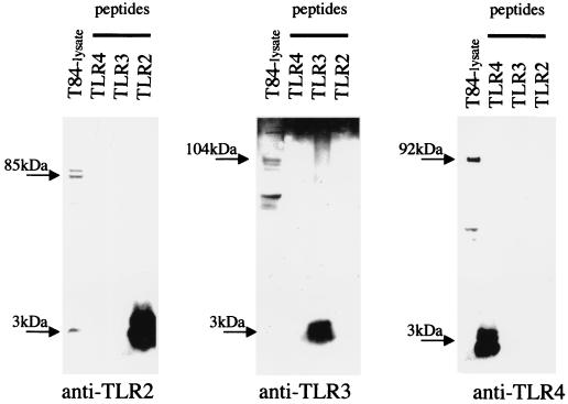
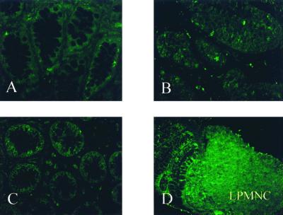
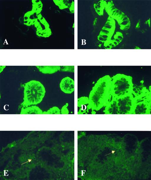
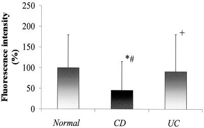
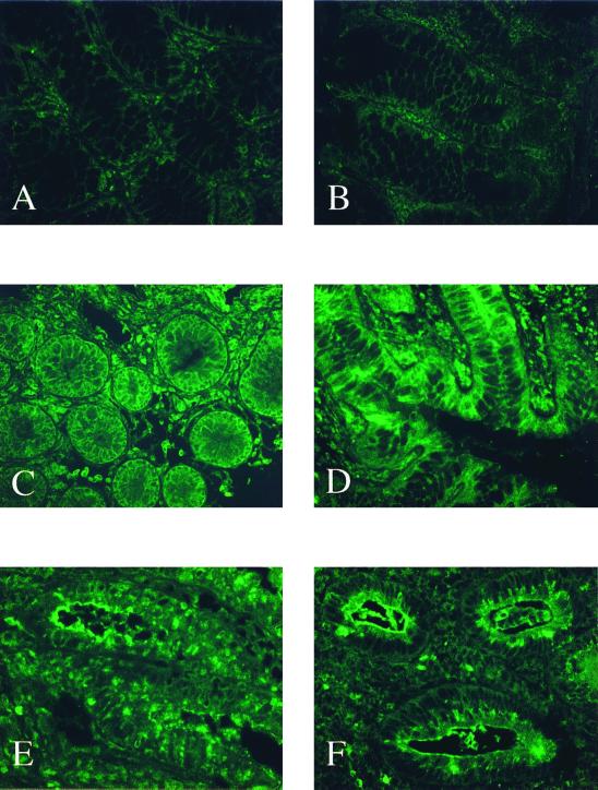
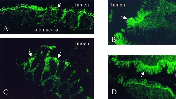
Initiation and perpetuation of the inflammatory intestinal responses in inflammatory bowel disease (IBD) may result from an exaggerated host defense reaction of the intestinal epithelium to endogenous lumenal bacterial flora. Intestinal epithelial cell lines constitutively express several functional Toll-like receptors (TLRs) which appear to be key regulators of the innate response system. The aim of this study was to characterize the expression pattern of TLR2, TLR3, TLR4, and TLR5 in primary intestinal epithelial cells from patients with IBD. Small intestinal and colonic biopsy specimens were collected from patients with IBD (Crohn's disease [CD], ulcerative colitis [UC]) and controls. Non-IBD specimens were assessed by immunofluorescence histochemistry using polyclonal antibodies specific for TLR2, TLR3, TLR4, and TLR5. Primary intestinal epithelial cells (IEC) of normal mucosa constitutively expressed TLR3 and TLR5, while TLR2 and TLR4 were only barely detectable. In active IBD, the expression of TLR3 and TLR4 was differentially modulated in the intestinal epithelium. TLR3 was significantly downregulated in IEC in active CD but not in UC. In contrast, TLR4 was strongly upregulated in both UC and CD. TLR2 and TLR5 expression remained unchanged in IBD. These data suggest that IBD may be associated with distinctive changes in selective TLR expression in the intestinal epithelium, implying that alterations in the innate response system may contribute to the pathogenesis of these disorders.
Figures
References
-
- Akashi S, Ogata H, Kirikae F, Kirikae T, Kawasaki K, Nishijima M, Shimazu R, Nagai Y, Fukudome K, Kimoto M, et al. Regulatory roles for CD14 and phosphatidylinositol in the signaling via toll-like receptor 4-MD-2. Biochem Biophys Res Commun. 2000;268:172–177. - PubMed
-
- Awane M, Andres P G, Li D J, Reinecker H C. NF-kB-inducing kinase is a common mediator of IL-17-, TNFa-, and IL-1β-induced chemokine promoter activation in intestinal epithelial cells. J Immunol. 1999;162:5337–5344. - PubMed
-
- Beutler B. Tlr4: central component of the sole mammalian LPS sensor. Curr Opin Immunol. 2000;12:20–26. - PubMed
-
- Bhan A, Mizoguchi E, Smith R N, Mizoguchi A. Colitis in transgenic and knockout animals as models of human inflammatory bowel disease. Immunol Rev. 1999;169:195–207. - PubMed
-
- Cario E, Rosenberg I M, Brandwein S L, Beck P L, Reinecker H C, Podolsky D K. Lipopolysaccharide activates distinct signaling pathways in intestinal epithelial cell lines expressing Toll-like receptors. J Immunol. 2000;164:966–972. - PubMed
Publication types
MeSH terms
Substances
Grants and funding
LinkOut - more resources
Full Text Sources
Other Literature Sources
