. 2000 Dec;68(12):7049-60.
doi: 10.1128/IAI.68.12.7049-7060.2000.

Human antibodies against a purified glucosylceramide from Cryptococcus neoformans inhibit cell budding and fungal growth

Affiliations

Human antibodies against a purified glucosylceramide from Cryptococcus neoformans inhibit cell budding and fungal growth

M L Rodrigues et al. Infect Immun. 2000 Dec.

Abstract

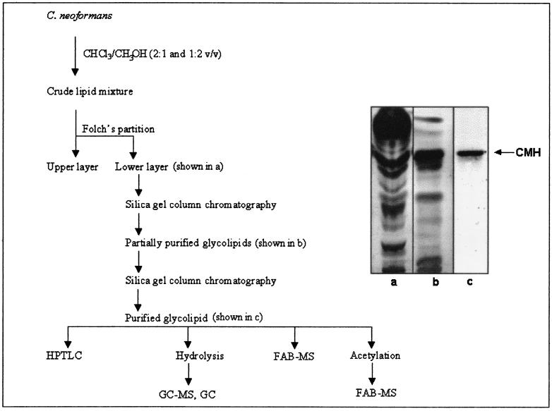
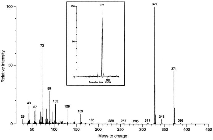
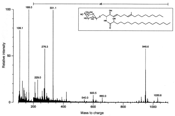
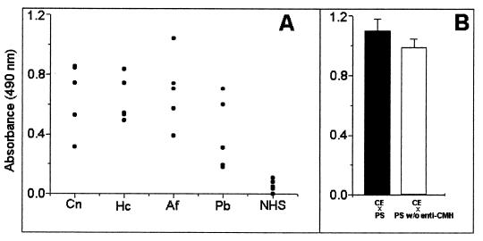
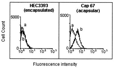
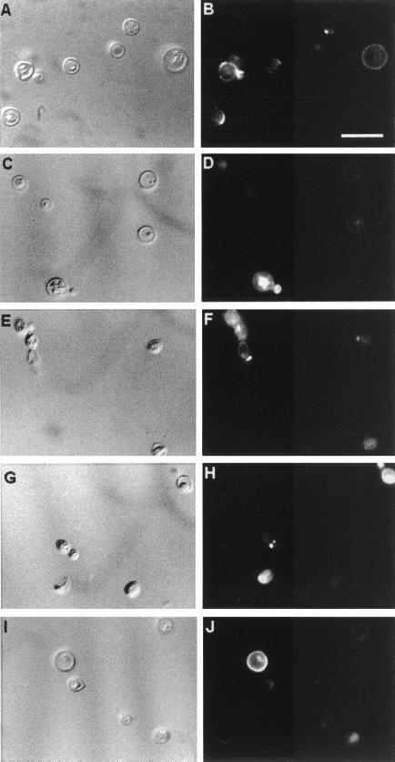
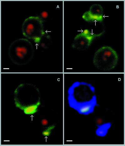
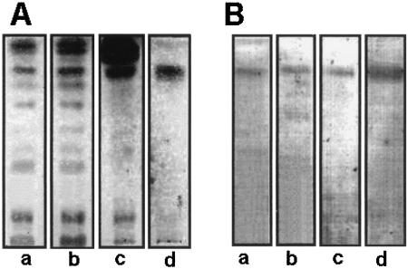
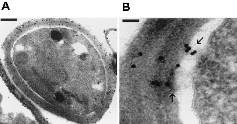
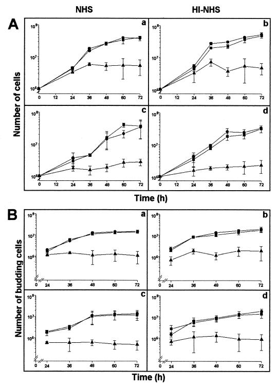
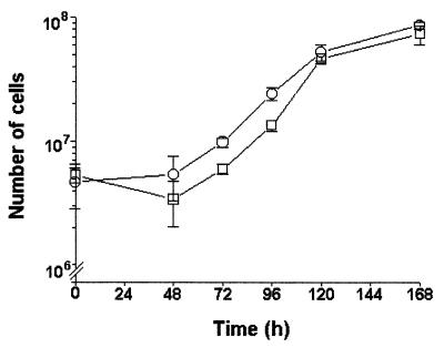
A major ceramide monohexoside (CMH) was purified from lipidic extracts of Cryptococcus neoformans. This molecule was analyzed by high-performance thin-layer chromatography (HPTLC), gas chromatography coupled with mass spectrometry, and fast atom bombardment-mass spectrometry. The cryptococcal CMH is a beta-glucosylceramide, with the carbohydrate residue attached to 9-methyl-4,8-sphingadienine in amidic linkage to 2-hydroxyoctadecanoic acid. Sera from patients with cryptococcosis and a few other mycoses reacted with the cryptococcal CMH. Specific antibodies were purified from patients' sera by immunoadsorption on the purified glycolipid followed by protein G affinity chromatography. The purified antibodies to CMH (mainly immunoglobulin G1) bound to different strains and serological types of C. neoformans, as shown by flow cytofluorimetry and immunofluorescence labeling. Transmission electron microscopy of yeasts labeled with immunogold-antibodies to CMH and immunostaining of isolated cell wall lipid extracts separated by HPTLC showed that the cryptococcal CMH predominantly localizes to the fungal cell wall. Confocal microscopy revealed that the beta-glucosylceramide accumulates mostly at the budding sites of dividing cells with a more disperse distribution at the cell surface of nondividing cells. The increased density of sphingolipid molecules seems to correlate with thickening of the cell wall, hence with its biosynthesis. The addition of human antibodies to CMH to cryptococcal cultures of both acapsular and encapsulated strains of C. neoformans inhibited cell budding and cell growth. This process was complement-independent and reversible upon removal of the antibodies. The present data suggest that the cryptococcal beta-glucosylceramide is a fungal antigen that plays a role on the cell wall synthesis and yeast budding and that antibodies raised against this component are inhibitory in vitro.


Figures

FIG. 1
FIG. 1
Isolation and purification of C. neoformans glucosylceramide. Steps of purification (left) and their corresponding fractions (right) are shown. Abbreviations are as defined in the text.
FIG. 2
FIG. 2
GC-MS analysis of the fatty acid moiety obtained from hydrolysis of the cryptococcal glucosylceramide. A single peak corresponding to a hydroxylated fatty acid was detected (inset), identified as 2-hydroxy-octadecanoic acid. For interpretation of the fragmentation, see Results.
FIG. 3
FIG. 3
FAB-MS of the peracetylated glucosylceramide from C. neoformans and its corresponding native structure. For interpretation of fragmentation, see Results.
FIG. 4
FIG. 4
(A) Reactivity of CMH with patients' antibodies. Sera from individuals with cryptococcosis (Cn), histoplasmosis (Hc), aspergillosis (Af), and paracoccidioidomycosis (Pb) recognize the cryptococcal CMH, while sera from normal individuals (NHS) do not. (B) Reactivity of crude extracts (CE) of C. neoformans with a pool of patients' serum (■) and patients' serum depleted of antibodies to CMH (□).
FIG. 5
FIG. 5
Flow cytometric analysis of encapsulated (HEC3393) and acapsular (cap 67) C. neoformans yeast cells incubated with antiglucosylceramide antibodies. Data curves: a, control cells (no antiglucosylceramide antibodies were added prior to incubation with FITC-labeled anti-human IgG); b, incubation of C. neoformans with antiglucosylceramide antibodies. After 1 h of exposure, the acapsular but not the encapsulated cells were reactive.
FIG. 6
FIG. 6
Surface distribution of the cryptococcal glucosylceramide. Acapsular (A and B) and encapsulated (C to J) C. neoformans yeast cells were incubated in the presence of antiglucosylceramide antibodies, followed by incubation with FITC-labeled anti-human IgG and observed in a fluorescence microscope. In control systems, in which no antiglucosylceramide antibodies were added prior to incubation with FITC-labeled anti-human IgG, no detectable fluorescence was observed (not shown). The left panels show C. neoformans yeasts using differential interferential contrast microscopy. Panels: A and B, acapsular cells; C and D, cryptococci of serotype A; E and F, serotype B cells; G and H, serotype C cells; and I and J, serotype D cells. Bar, 10 μm.
FIG. 7
FIG. 7
Confocal microscopy of CMH at the cell surface of acapsular C. neoformans cells. (A to C) Budding cells show the CMH (arrows), mainly concentrated at the budding region, while mature nondividing cells have some weakly labeled clusters on the cell surface. (D) Cells shown in Fig. 7C were analyzed by overlaying the specific fluorescent reaction of surface CMH with antibodies to them (arrows) and the autofluorescence of the cryptococcal cell wall (blue). This technique shows that the polar concentration of CMH colocalizes with a thickened cell wall to the site of bud formation. Autofluorescence of the nucleus is shown in red. Bars, 1 μm.
FIG. 8
FIG. 8
HPTLC (A) and HPTLC-immunostaining (B) of glycolipids obtained from serotype B (lanes a) and D (lanes b) cells or from cell wall preparations from cap 67 cells (lanes c) compared with the purified glucosylceramide from HEC3393 cells (lanes d). Orcinol-reactive bands with Rf values similar to that of the purified cryptococcal glucosylceramide were detected by HPTLC. Immunostaining showed that antibodies to CMH recognized components with migration similar to that of CMH.
FIG. 9
FIG. 9
Cryptococcal CMH are located at the fungal cell wall. (A) Transmission electron microscopy showed an extensive binding of antibodies to CMH to the cryptococcal cell wall, integrated or detached from the cells. Bar, 0.5 μm. (B) Possible CMH-containing vesicles are seen (arrows), which can move across the periplasmic space and deposit cell membrane constituents on the cell wall. Bar, 0.1 μm.
FIG. 10
FIG. 10
Influence of antibodies to CMH on the cell growth (A) and budding (B) of encapsular (a and b) and acapsulated (c and d) C. neoformans. The culture medium was supplemented with NHS (a and c) or heat-inactivated NHS (HI-NHS, b and d). Addition of antibodies to CMH to the cell cultures at 10 μg/ml (▴) inhibited fungal growth and budding in both acapsular and encapsulated cells. ●, Untreated cells; ■, cells treated with normal human IgG, processed as for the antibodies to CMH, at 100 μg/ml.
FIG. 11
FIG. 11
Antibodies to CMH are fungistatic but not fungicidal for C. neoformans. Replacement of the antibody-containing medium by BHI restored the fungal growth of encapsulated (strain HEC3393) cells previously cultivated for 48 h in BHI with antibodies to CMH in normal (○) or heat-inactivated NHS (□).

References

    1. Barreto-Bergter E, Vermelho A B, Hartmann R, Pohlentz G, Klein R A, Egge H. Structural characterization of neutral glycosphingolipids from Trypanosoma cruzi. Mol Biochem Parasitol. 1992;51:263–270. - PubMed
    1. Barreto-Bergter E, Travassos L R, Gorin P A J. Chemical structure of the d-galacto-d-mannan component from hyphae of Aspergillus niger and other Aspergillus spp. Carbohydr Res. 1980;86:273–285.
    1. Bielawska A, Linardic C M, Hannun Y A. Modulation of cell growth and differentiation by ceramide. FEBS Lett. 1992;307:211–214. - PubMed
    1. Chen L C, Blank E S, Casadevall A. Extracellular proteinase activity of Cryptococcus neoformans. Clin Diagn Lab Immunol. 1996;3:570–574. - PMC - PubMed
    1. Chen S C A, Muller M, Zhou J Z, Wright L C, Sorrel T C. Phospholipase activity in Cryptococcus neoformans: a new virulence factor? J Infect Dis. 1997;175:414–420. - PubMed

Publication types

LinkOut - more resources