. 2000 Dec;68(12):7114-21.
doi: 10.1128/IAI.68.12.7114-7121.2000.

Surface protein variation by expression site switching in the relapsing fever agent Borrelia hermsii

Affiliations

Surface protein variation by expression site switching in the relapsing fever agent Borrelia hermsii

A G Barbour et al. Infect Immun. 2000 Dec.

Abstract

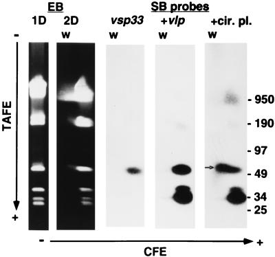
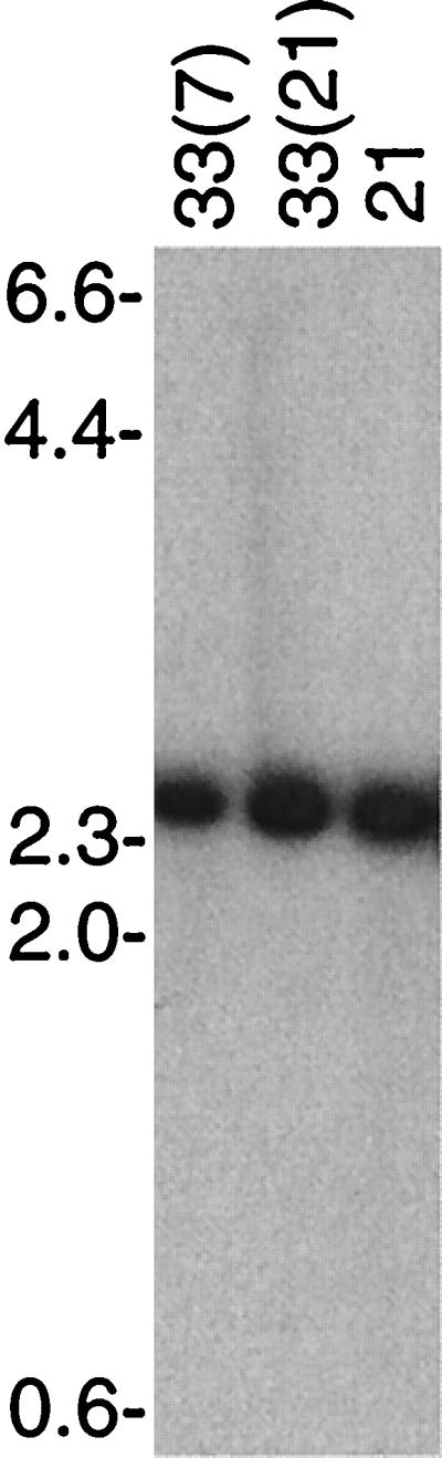
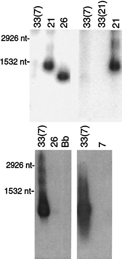
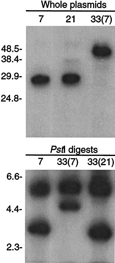
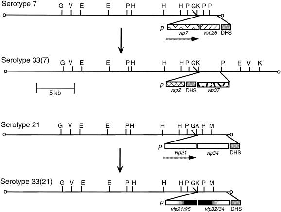
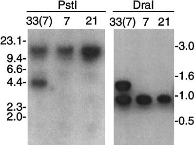
Borrelia hermsii, an agent of relapsing fever, undergoes antigenic variation of serotype-specifying membrane proteins during mammalian infections. When B. hermsii is cultivated in broth medium, one serotype, 33, eventually predominates in the population. Serotype 33 has also been found to be dominant in ticks but not in mammalian hosts. We investigated the biology and genetics of two independently derived clonal populations of serotype 33 of B. hermsii. Both isolates infected immunodeficient mice, but serotype 33 cells were limited in number and were only transiently present in the blood. Probes for vsp33, which encodes the serotype-specifying Vsp33 outer membrane protein, revealed that the gene was located on a 53-kb linear plasmid and that there was only one locus for the gene in serotype 33. The vsp33 probe and probes for other variable membrane protein genes showed that expression of Vsp33 was determined at the level of transcription and that when the vsp33 expression site was active, an expression site for other variable proteins was silent. The study confirmed that serotype 33 is distinct from other serotypes of B. hermsii in its biology and demonstrated that B. hermsii can change its major surface protein through switching between two expression sites.


Figures

FIG. 1
FIG. 1
One-dimensional (1D) and two-dimensional (2D) gels with ethidium bromide (EB) staining and Southern blot (SB) analysis of a two-dimensional gel of total DNA of serotype 33 of B. hermsii strain HS1. The blot was sequentially hybridized first with probe 33-P for vsp33, then with probe 7-P for vlp7 (+vlp), and finally with probe CP for the circular plasmid (+cir. pl.). For the two-dimensional gel in 1.0% agarose, electrophoresis was first transverse alternating field (TAFE) and then constant field (CFE). The long arrows show the direction of migration from cathode (−) to anode (+). The migrations of linear molecular size standards (in kilobases) in the TAFE dimension are shown on the right. The small arrow indicates the position of the circular plasmid behind the 53-kb linear plasmid by CFE. The conditions for TAFE at 181 mA in 0.5× Tris-borate-EDTA buffer were a 4-s pulse for 30 min, 1-s pulse for 9 h, and 5-s pulse for 9 h.
FIG. 2
FIG. 2
Southern blot analysis of HindIII-digested total DNA from serotypes 33(7), 33(21), and 21 of B. hermsii that was probed with probe 33-P for vsp33. The agarose concentration was 0.7%, and the locations of the migrations of molecular size markers (in kilobases) are shown on the left.
FIG. 3
FIG. 3
The vsp33 gene is transcribed in serotype 33 but not in other serotypes. Northern blot analyses of RNA of serotypes 33(7) and 33(21) of B. hermsii and of B. burgdorferi (Bb) with probes specific for vsp33 (above) or with an oligonucleotide (V-O) that binds to the 5′ end of all other known vsp and vlp genes (below) were carried out. Formaldehyde-denatured total RNA was separated by electrophoresis in 1.5% agarose. (Above) Two separate experiments are shown: 33(7), 21, and 26 on the left and 33(7), 33(21), and 21 on the right. (Below) Two separate experiments are shown: 33(7), 26, and B. burgdorferi (Bb) on the left and 33(7) and 7 on the right. The probe for the left lower blot was 33-O, and the probe for the right lower blot was 33-P. The sizes of 23S (2,926 nucleotides [nt]) and 16S (1,532 nt) of rRNAs of Borrelia are indicated to the left (39).
FIG. 4
FIG. 4
Southern blot analysis of intact plasmids of serotypes 7, 21, and 33(7) (above) and PstI digests of DNA from serotypes 7, 33(7), and 33(21) (below) with ES1 probes. For the upper blot, the field inversion electrophoresis gel had 1.0% agarose and the p7.41 probe was used. For the lower blot, the constant field electrophoresis gel had 0.7% agarose and the pE21 probe, which includes a PstI site, was used. The migrations of molecular size standards (in kilobases) are shown on the left.
FIG. 5
FIG. 5
Partial physical maps of expression site plasmids before and after an in vitro switch to serotype 33 from serotype 7 (above) or from serotype 21 (below). For each switch, the changes in restriction maps for the linear plasmid bearing the telomeric expression site for either vlp7 or vlp21 in serotypes 7 or 21, respectively, are shown. The regions containing the putative promoter (p), the vsp or vlp genes, and the repetitive DHS sequence (25) are expanded below each plasmid. The different sequences are indicated by different patterns, such as the cross-hatched pattern for vsp26. The exception is the representation of vlp21 and vlp34 with the same pattern in the lower panel even though they have different sequences. For serotype 33(21), the chimeric vlp genes are indicated by the white-black gradients. Genes that were expressed, vlp7 in serotype 7 and vlp21 in serotype 21, are indicated by the horizontal arrow beneath. The restriction enzymes were BglII (G), EcoRV (V), EcoRI (E), PstI (P), HindIII, KpnI (K), and MspI (M). The restriction maps were determined using plasmid and oligonucleotide probes specific for the ES1 promoter and its 5′ flanking region, as well as oligonucleotide probes specific for vsp2, vlp7, vlp21, and vlp25.
FIG. 6
FIG. 6
Southern blot analysis of PstI- or DraI-digested total DNA of serotypes 33(7), 7, and 21 that was hybridized with probe 2-O for vsp2. The agarose concentrations were 0.7% for PstI digests and 1.0% for DraI digests. The migrations of molecular size standards (in kilobases) are shown.

References

    1. Barbour A G. Antigenic variation of a relapsing fever Borrelia species. Annu Rev Microbiol. 1990;44:155–171. - PubMed
    1. Barbour A G. Isolation and cultivation of Lyme disease spirochetes. Yale J Biol Med. 1984;57:521–525. - PMC - PubMed
    1. Barbour A G. Linear DNA of Borrelia species and antigenic variation. Trends Microbiol. 1993;1:236–239. - PubMed
    1. Barbour A G, Barrera O, Judd R C. Structural analysis of the variable major proteins of Borrelia hermsii. J Exp Med. 1983;158:2127–2140. - PMC - PubMed
    1. Barbour A G, Burman N, Carter C J, Kitten T, Bergstrom S. Variable antigen genes of the relapsing fever agent Borrelia hermsii are activated by promoter addition. Mol Microbiol. 1991;5:489–493. - PubMed

Publication types

Substances

LinkOut - more resources