. 2000 Dec;68(12):7202-8.
doi: 10.1128/IAI.68.12.7202-7208.2000.

Localization of dysfunctional tight junctions in Salmonella enterica serovar typhimurium-infected epithelial layers

Affiliations

Localization of dysfunctional tight junctions in Salmonella enterica serovar typhimurium-infected epithelial layers

M A Jepson et al. Infect Immun. 2000 Dec.

Abstract

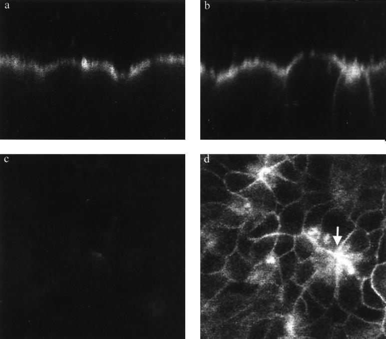
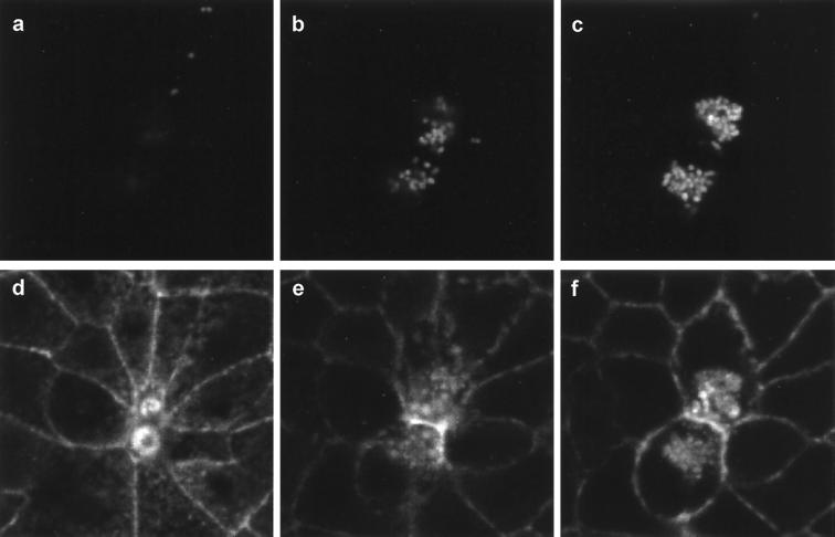
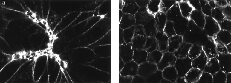
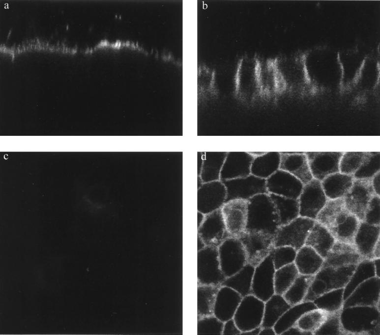
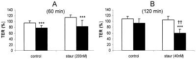
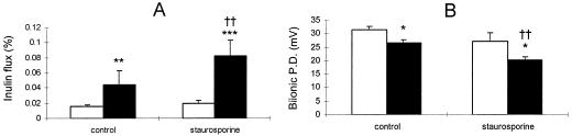
Infection of polarized MDCK epithelial layers by Salmonella enterica serovar Typhimurium is accompanied by increased tight junction permeability and by contraction of perijunctional actinomyosin. We localized dysfunctional tight junctions in serovar Typhimurium-infected MDCK layers by imaging apical-basolateral intramembrane diffusion of fluorescent lipid and found that loss of the apical-basolateral diffusion barrier (tight junction fence function) was most marked in areas of prominent perijunctional contraction. The protein kinase inhibitor staurosporine prevented perijunctional contraction but did not reverse the effects of serovar Typhimurium on tight junction barrier function. Hence, perijunctional contraction is not required for Salmonella-induced tight junction dysfunction and this epithelial response to infection may be multifactorial.


Figures

FIG. 1
FIG. 1
Serovar Typhimurium infection causes disruption of the apical-basolateral intramembrane diffusion barrier function of tight junctions in MDCK epithelia. Confocal imaging of BODIPY-sphingomyelin-labeled MDCK epithelia demonstrates retention of fluorescence in the apical membrane of control cell layers, as revealed in an xz (transverse) image (a). Serovar Typhimurium infection for 60 min causes a reduction in the ability of some of the tight junctions to limit apical-basolateral intramembrane diffusion of BODIPY-sphingomyelin, resulting in the appearance of variable mounts of the tracer throughout the lateral membranes of some cells (xz image) (b). A relationship between perijunctional contraction and tight junction dysfunction is revealed in a confocal optical section 10 to 12 μm below the apical surface of the cell layer (d), which reveals relatively high concentrations of dye in the lateral membranes of cells which are contracted as a result of infection (arrow). A similar optical section of uninfected cell layers 10 to 12 μm below the apical surface confirms that there is minimal diffusion of the lipid across tight junctions (c), since little fluorescence is detected in the lateral membranes. All images were obtained under identical imaging parameters with a Leica TCS NT confocal system attached to a Leica DM RBE epifluorescence microscope and equipped with a Kr-Ar mixed-gas laser. Magnification, ×1,010.
FIG. 2
FIG. 2
Perijunctional contraction is associated with serovar Typhimurium invasion. MDCK monolayers were infected for 60 min with serovar Typhimurium expressing green fluorescent protein (GFP) and then fixed and stained with phalloidin-TRITC to localize F-actin. Dual-channel confocal imaging reveals GFP-expressing bacteria (a to c) and F-actin (d to f) at varying depths within the infected cell layer 2 μm (b,e) and 4 μm (c,f) below the optical section at the apex of the cells (a,d). Images were acquired with a Leica TCS NT confocal system attached to a Leica DM RBE epifluorescence microscope and equipped with a Kr-Ar mixed-gas laser. Note that actin accumulation in the vicinity of invaded bacteria and a small amount of bleedthrough of GFP fluorescence into the TRITC channel allow location of Salmonella (d to f). An optical section at the apex of the cell layer reveals two regions of actin accumulation (corresponding to membrane ruffles on cells) which are contracted (note also the marked stretching of surrounding cells). Optical sections 2 μm (b,e) and 4 μm (c,f) below the apical pole reveal that these two contracted cells have typical profiles beneath the level of the perijunctional actinomyosin ring and have been invaded by several GFP-expressing serovar Typhimurium bacteria. Magnification, ×1,300.
FIG. 3
FIG. 3
Staurosporine inhibits serovar Typhimurium-induced perijunctional actinomyosin contraction. Phalloidin-TRITC staining of MDCK monolayers infected with serovar Typhimurium SL1344 for 60 min without staurosporine treatment (a) or after pretreatment with 200 nM staurosporine (b). Confocal images obtained in the xy plane reveals extensive distortion of MDCK cells after 60 min of infection in the absence of staurosporine (a). Contracted cells are intensely stained with phalloidin-TRITC, and neighboring cells exhibit clear distortion due to the stretching imposed by contraction of infected cells. MDCK monolayers infected with serovar Typhimurium treated with staurosporine (b) reveal a regular cell shape due to the absence of contraction of the perijunctional actin ring. Images were acquired with a Leica TCS NT confocal system attached to a Leica DM RBE epifluorescence microscope and equipped with a Kr-Ar mixed-gas laser. Magnification, ×640.
FIG. 4
FIG. 4
Serovar Typhimurium-induced disruption of the apical-basolateral intramembrane diffusion barrier in the presence of staurosporine. Confocal imaging of BODIPY-sphingomyelin labeling reveals the retention of fluorescence in the apical membrane of 200 nM staurosporine-treated uninfected MDCK layers, as revealed in an xz (transverse) image (a). Infection of staurosporine-treated cells with serovar Typhimurium for 60 min impairs the apical-basolateral intramembrane diffusion barrier, resulting in the appearance of BODIPY-sphingomyelin throughout the lateral membranes of all cells (xz image) (b). Cells infected with serovar Typhimurium in the presence of staurosporine do not exhibit distortion that is due to perijunctional contraction but do accumulate BODIPY-sphingomyelin in lateral membranes throughout the cell layer, revealed by xy optical sectioning 8 to 10 μm below the apical surface (d; compare with Fig. 1d). A similar optical section of uninfected, staurosporine-treated cell layers reveals minimal diffusion of the lipid across tight junctions (c). All images were obtained under identical imaging parameters with a Leica TCS NT confocal system attached to a Leica DM RBE epifluorescence microscope and equipped with a Kr-Ar mixed-gas laser. Magnification, ×1,010.
FIG. 5
FIG. 5
Serovar Typhimurium induces a decrease in TER which is not reversed by staurosporine. MDCK layers were infected with serovar Typhimurium at an MOI of 100 following treatment with 200 nM (A) or 40 nM (B) staurosporine (or equivalent concentrations of the carrier, dimethyl sulfoxide). Tight junction permeability was then assessed over a 60-min (A) or 120-min (B) time course, and a comparison was made between uninfected layers (empty bars) and Salmonella-infected layers (solid bars). Staurosporine (200 nM) fails to reverse the drop in TER induced during 60 min of serovar Typhimurium infection (A). Decreasing the staurosporine concentration to 40 nM allowed the experiment to be prolonged while preventing any effect of staurosporine itself on TER. Under these conditions staurosporine enhances the effect of Salmonella on TER (B). All data are expressed as means ± standard deviations (n = 10 to 12 for panel A and n = 4 for panel B). The statistical significance of differences was assessed by Student's unpaired t test with significance set at P values of <0.05. Significant differences between uninfected and infected layers are indicated by three asterisks (P < 0.001), while significant differences between staurosporine-treated and control layers are indicated by double daggers (P < 0.01).
FIG. 6
FIG. 6
Staurosporine treatment enhances the effects of serovar Typhimurium infection on tight junction permeability. MDCK layers were infected with serovar Typhimurium at an MOI of 100 following treatment with 200 nM staurosporine (or equivalent concentrations of the carrier, dimethyl sulfoxide). Tight junction permeability was then assessed over a 30-min time course, and a comparison was made between uninfected layers (empty bars) and Salmonella-infected layers (solid bars). Inulin flux (A) measured by the appearance of apically applied [14C]inulin in the basal bathing medium was significantly increased by Salmonella infection and further enhanced by staurosporine. In separate experiments (B), bi-ionic potential difference (P.D.) (measured following isoosmotic replacement of sodium in the basal medium with choline [8]) was significantly decreased by serovar Typhimurium infection and further diminished by staurosporine treatment. All data are expressed as means ± standard deviations (n = 6 for panel A and n = 3 for panel B). The statistical significance of differences was assessed by Student's unpaired t test with significance set at P values of < 0.05. Significant differences between uninfected and infected layers are indicated by asterisks (∗, P < 0.05; ∗∗, P < 0.01; ∗∗∗, P < 0.001), while significant differences between staurosporine-treated and control layers are indicated by double daggers (P < 0.01).

Similar articles

Cited by

References

    1. Azghani A O, Gray L D, Johnson A R. A bacterial protease perturbs the paracellular barrier function of transporting epithelial monolayers in culture. Infect Immun. 1993;61:2681–2686. - PMC - PubMed
    1. Bain C, Keller R, Collington G K, Trabulsi L R, Knutton S. Increased levels of intracellular calcium are not required for the formation of attaching and effacing lesions by enteropathogenic and enterohemorrhagic Escherichia coli. Infect Immun. 1998;66:3900–3908. - PMC - PubMed
    1. Balda M S, Whitney J A, Flores C, González S, Cereijido M, Matter K. Functional dissociation of paracellular permeability and transepithelial electrical resistance and disruption of the apical-basolateral intramembrane diffusion barrier by expression of a mutant tight junction membrane protein. J Cell Biol. 1996;134:1031–1049. - PMC - PubMed
    1. Brumell J H, Steele-Mortimer O, Finlay B B. Bacterial invasion: force feeding by Salmonella. Curr Biol. 1999;9:R277–R280. - PubMed
    1. Citi S. Protein kinase inhibitors prevent junction dissociation induced by low extracellular calcium in MDCK epithelial cells. J Biol Chem. 1992;117:169–178. - PMC - PubMed

Publication types

LinkOut - more resources