. 2000 Nov 20;192(10):1425-40.
doi: 10.1084/jem.192.10.1425.

Lymph-borne chemokines and other low molecular weight molecules reach high endothelial venules via specialized conduits while a functional barrier limits access to the lymphocyte microenvironments in lymph node cortex

Affiliations

Lymph-borne chemokines and other low molecular weight molecules reach high endothelial venules via specialized conduits while a functional barrier limits access to the lymphocyte microenvironments in lymph node cortex

J E Gretz et al. J Exp Med. .

Abstract

Lymph-borne, soluble factors (e.g., chemokines and others) influence lymphocyte recirculation and endothelial phenotype at high endothelial venules (HEVs) in lymph node cortex. Yet the route lymph-borne soluble molecules travel from the subcapsular sinus to the HEVs is unclear. Therefore, we injected subcutaneously into mice and rats a wide variety of fluorophore-labeled, soluble molecules and examined their distribution in the draining lymph nodes. Rather than percolating throughout the draining lymph node, all molecules, including microbial lipopolysaccharide, were very visible in the subcapsular and medullary sinuses but were largely excluded from the cortical lymphocyte microenvironments. Exclusion prevailed even during the acute lymph node enlargement accompanying viral infection. However, low molecular mass (MW) molecules, including chemokines, did gain entry into the cortex, but in a very defined manner. Low MW, fluorophore-labeled molecules highlighted the subcapsular sinus, the reticular fibers, and the abluminal and luminal surfaces of the associated HEVs. These low MW molecules were in the fibers of the reticular network, a meshwork of collagen fibers ensheathed by fibroblastic reticular cells that connects the subcapsular sinus floor and the HEVs by intertwining with their basement membranes. Thus, low MW, lymph-borne molecules, including chemokines, traveled rapidly from the subcapsular sinus to the HEVs using the reticular network as a conduit.


Figures

Figure 1
Figure 1
A model of movement of lymph-borne, soluble molecules through the lymph node; distribution is dependent on molecular size. Rather than percolating through the cortical parenchyma, soluble molecules of both high and low MW moved through the lymph node via the subcapsular and medullary sinuses (note that cortical sinuses are not pictured here). (A) Although high MW molecules were excluded from lymph node cortex and moved through the lymph node only via the sinuses, rapid movement of low MW molecules (B) from the subcapsular sinus to the HEVs was restricted to the fibers of the reticular network. (C) Comparison of the distribution in the lymph node of lymph-borne soluble molecules by size. SCS, subcapsular sinus; T, T cell area; B, B cell area, follicle; RF, reticular fiber; FRC, fibroblastic reticular cell; G, HEV endothelial glycocalyx; M, medulla.
Figure 3
Figure 3
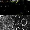
Low MW, soluble molecules enter lymph node cortex and distribute in a pattern resembling reticular network. Distribution of lymph-borne, fluorophore-labeled dextrans in draining rat and mouse lymph nodes is compared with Gomori silver staining of the reticular network in a mouse lymph node (confocal and bright-field micrographs, respectively). After the subcutaneous injection of dextrans, draining lymph nodes were excised and immersion fixed within 10–12 min. Lymph node sections are 10 μm thick. Subcapsular sinuses (between arrows) and typical HEVs (arrowheads) are labeled. (A) Fluorescein-labeled dextran (3 kD, green) and Texas red–labeled dextran (70 kD, red). (B) Fluorescein-labeled dextran (40 kD, green) and Texas red–labeled dextran (70 kD, red). (C) Gomori reticulin stain of mouse lymph node. (D) Higher magnification of HEVs in rat lymph node after subcutaneous injection of Texas red–labeled dextran (10 kD, white). Note the dextran in lumen (arrow). Bars, (A–C) 50 μm; (D) 25 μm.
Figure 2
Figure 2
Exclusion of high MW molecules and LPS from lymph node cortex. Confocal micrographs showing subcapsular sinuses (between arrows) and underlying cortices of draining mouse lymph nodes after fluorophore-labeled molecules were injected subcutaneously. The lymph nodes were excised and immersion fixed within 10–12 min after injection. Lymph node sections, 10 μm thick. Bars, 50 μm. (A) Fluorescein-labeled dextran (500 kD, green) and Texas red–labeled dextran (2,000 kD, red) in a mouse axillary lymph node after footpad and intravenous injections, respectively. (B) Anti–mouse MHC class I–FITC (150 kD, white) in mouse lymph node. Inset shows control node; note the autofluorescent cells in both control and experimental conditions due to gain used (C). LPS-FITC (white) in mouse lymph node. Control node in inset.
Figure 4
Figure 4
Typical protein immunogens have restricted distribution in cortices of draining lymph nodes after subcutaneous injection. Confocal micrographs of lymph node sections (10 μm) show FITC-labeled protein in subcapsular sinus (between arrows), highlighting reticular network and HEVs (arrowheads). Bars, 50 μm. (A) OVA-FITC (white) in the draining axillary lymph node. Lymph node was excised and fixed 15 min after injection of OVA into the mouse footpad. (B) HEL-FITC (green) in draining mouse lymph node. Lymph node was excised and fixed 5 min after injection of HEL into the footpad. Lymph node section was labeled topically with Cy-Chrome–labeled anti–mouse CD45R/B220 antibody to visualize B cells (red).
Figure 6
Figure 6
Lymph-borne, low MW molecules are in reticular fibers in the draining lymph nodes. (A) Confocal microscopy image of a draining rat mesenteric lymph node after the injection of Texas red–labeled dextran (10 kD, red) into the associated Peyer's patch. Reticular fibers (red) descend vertically from the dextran-filled subcapsular sinus (arrows). Topical application of the lymph node section with WGA-FITC (green) highlights cells associated with reticular network. A capillary intertwined by the reticular network runs horizontally across the image (arrowheads). Bar, 18 μm. (B) Optical cross-section of two reticular fibers at the white line in A. Note the Texas red–dextran encircled by WGA-FITC. Bar, 4 μm. (C and D) TEMs of reticular fibers ensheathed by fibroblastic reticular cells (arrowheads) in rat mesenteric lymph nodes. L, the nuclei of cortical lymphocytes. In C, the reticular fiber is filled with lymph-borne HRP; D is from the control of the same experiment.
Figure 5
Figure 5
Lymph-borne molecules travel unidirectionally from the subcapsular sinus (arrows) through the reticular network toward HEVs (small arrowheads) in the lymph node cortex and are excluded from the lymphocyte microenvironments. Lymph node sections, 10 μm. Bars, 50 μm. (A) Lymphocytes, reticular fibers, and HEVs are visible in this confocal micrograph of a fresh-frozen section of mouse lymph node, topically labeled with FITC-labeled lectin, PSA (PSA-FITC). (B) Lymph-borne PSA-FITC highlights the subcapsular (arrows) and medullary (large arrowheads) sinuses and an incomplete reticular network starting at the subcapsular sinus. Small arrowheads point to three HEVs that are also highlighted by the lymph-borne PSA-FITC: two HEVs cut in cross-section (small arrowheads, middle right) and a single HEV with two branches, cut longitudinally (three small arrowheads, top left). Note the absence of labeled lymphocytes in the cortex. This draining lymph node was excised and fixed 30 min after injection of PSA-FITC into the footpad.
Figure 8
Figure 8
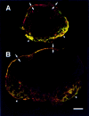
Despite acute changes in lymph node system during viral infection, patterns of distribution of lymph-borne, soluble molecules are preserved. Montages of confocal images of lymph node sections (10 μm) from control (A) and virally infected (B) draining mouse popliteal lymph nodes are presented here. Mouse popliteal lymph nodes were excised and immersion fixed 5 min after footpads were injected with fixable, fluorophore-labeled dextrans (10-kD dextran–Texas red [red], and 70-kD dextran–fluorescein [green]). Bar, 160 μm. (A) Control lymph node. (B) Virally infected lymph node, 8 h after footpad injection with modified vaccinia virus. Note that both low and high MW fluorophore-labeled dextrans filled the subcapsular (arrows) and medullary sinuses (arrowheads), whereas the lower MW dextran (red) highlights the reticular network and associated HEVs in the cortex of each lymph node.
Figure 7
Figure 7
Particulate accumulation of high MW tracer in cortex after, not during, the initial bolus of soluble tracer in subcapsular sinus. (A) High MW dextran only in subcapsular sinus (arrows) at 5 min after subcutaneous injection. (B) At 30 min, lower intensity signal of high MW dextran in subcapsular sinus (arrows) but high persistent signal in medulla (on right). (C) Control lymph node. (D) Montage of adjacent areas of popliteal lymph node. Focal accumulation of high MW dextran (2,000 kD, fluorescein labeled, green) at 4 h within parenchyma contrasts with reticular-type distribution of low MW dextran (10 kD, Texas red, red) at 10 min (subcapsular sinus, arrows; HEV, arrowheads). However, despite different times of administration, both tracers coaccumulated in a cellular-type distribution within a medullary sinus. Bars, 50 μm.
Figure 9
Figure 9
Lymph-borne chemokines travel to HEVs via reticular network “conduits.” Confocal images shown of lymph-borne MIP-1α–FITC in subcapsular sinus (arrows), cortical/medullary sinuses (large arrowheads), and reticular fibers descending from the subcapsular sinus floor to several HEVs (small arrowheads) in a draining mouse axillary lymph node. The lymph node was excised and immersion fixed 15 min after subcutaneous injection of the chemokine into the footpad. (A) Cortex. (B) Cortical/medullary sinuses (large arrowheads). (C) Control showing autofluorescent cells due to gain used. Bars, 50 μm.

References

    1. Gowans J.L., Knight E.J. The route of re-circulation of lymphocytes in rat. Proc. R. Soc. Lond. Ser. B. 1964;159:257–282. - PubMed
    1. Butcher E.C. Leukocyte-endothelial cell recognitionthree (or more) steps to specificity and diversity. Cell. 1991;67:1033–1036. - PubMed
    1. Shimizu Y., Newman W., Tanaka Y., Shaw S. Lymphocyte interactions with endothelial cells. Immunol. Today. 1992;13:106–112. - PubMed
    1. Tanaka Y., Adams D.H., Hubscher S., Hirano H., Siebenlist U., Shaw S. T-cell adhesion induced by proteoglycan-immobilized cytokine MIP-1 beta. Nature. 1993;361:79–82. - PubMed
    1. Tedla N., Palladinetti P., Kelly M., Kumar R.K., DiGirolamo N., Chattophadhay U., Cooke B., Truskett P., Dwyer J., Wakefield D., Lloyd A. Chemokines and T lymphocyte recruitment to lymph nodes in HIV infection. Am. J. Pathol. 1996;148:1367–1373. - PMC - PubMed

Publication types