. 2000 Nov 20;192(10):1515-20.
doi: 10.1084/jem.192.10.1515.

Requirement of the chemokine receptor CXCR3 for acute allograft rejection

Affiliations

Requirement of the chemokine receptor CXCR3 for acute allograft rejection

W W Hancock et al. J Exp Med. .

Abstract

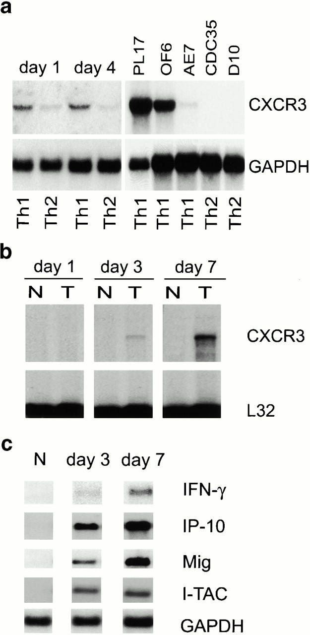
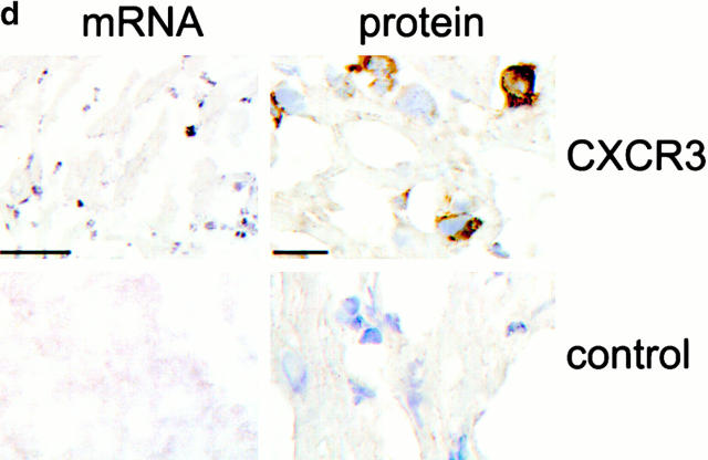
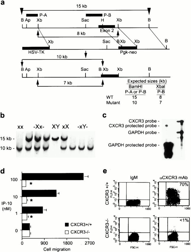
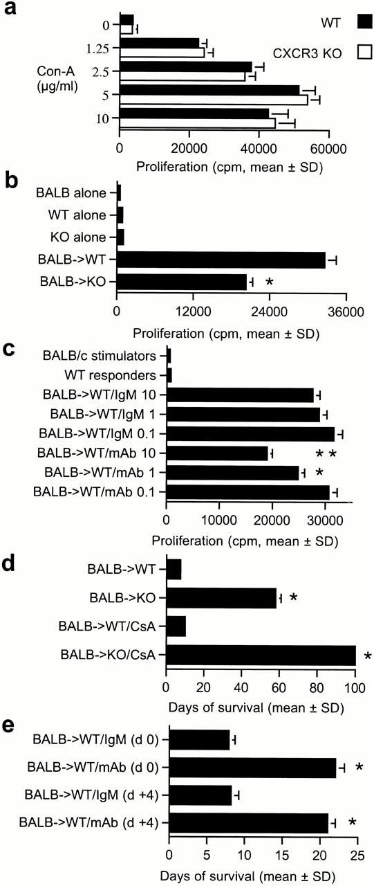
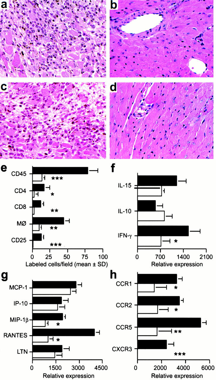
Chemokines provide signals for activation and recruitment of effector cells into sites of inflammation, acting via specific G protein-coupled receptors. However, in vitro data demonstrating the presence of multiple ligands for a given chemokine receptor, and often multiple receptors for a given chemokine, have led to concerns of biologic redundancy. Here we show that acute cardiac allograft rejection is accompanied by progressive intragraft production of the chemokines interferon (IFN)-gamma-inducible protein of 10 kD (IP-10), monokine induced by IFN-gamma (Mig), and IFN-inducible T cell alpha chemoattractant (I-TAC), and by infiltration of activated T cells bearing the corresponding chemokine receptor, CXCR3. We used three in vivo models to demonstrate a role for CXCR3 in the development of transplant rejection. First, CXCR3-deficient (CXCR3(-/)-) mice showed profound resistance to development of acute allograft rejection. Second, CXCR3(-/)- allograft recipients treated with a brief, subtherapeutic course of cyclosporin A maintained their allografts permanently and without evidence of chronic rejection. Third, CXCR(+/+) mice treated with an anti-CXCR3 monoclonal antibody showed prolongation of allograft survival, even if begun after the onset of rejection. Taken in conjunction with our findings of CXCR3 expression in rejecting human cardiac allografts, we conclude that CXCR3 plays a key role in T cell activation, recruitment, and allograft destruction.


Figures

Figure 1
Figure 1
Analysis of CXCR3 expression by activated T cells in vitro and during graft rejection. (a) Northern blot analysis of CXCR3 expression by primary T cells activated under polarizing conditions and by Th1 cell clones. (b) RNase protection assays of CXCR3 expression in serial cardiac transplants (T) showing rejection by day 7 but not corresponding native hearts (N). (c) Northern blots showing serial allograft but not native (N) heart expression of IFN-γ–induced CXCR3 ligands IP-10, Mig, and I-TAC. (d) Graft CXCR3 mRNA (blue, left) expression is restricted to focal round cells (sense control, bottom left); immunoperoxidase at high power (brown, right) shows CXCR3 protein expression by infiltrating mononuclear leukocytes (IgM control, bottom right). Bars, 100 and 10 μm, respectively.
Figure 1
Figure 1
Analysis of CXCR3 expression by activated T cells in vitro and during graft rejection. (a) Northern blot analysis of CXCR3 expression by primary T cells activated under polarizing conditions and by Th1 cell clones. (b) RNase protection assays of CXCR3 expression in serial cardiac transplants (T) showing rejection by day 7 but not corresponding native hearts (N). (c) Northern blots showing serial allograft but not native (N) heart expression of IFN-γ–induced CXCR3 ligands IP-10, Mig, and I-TAC. (d) Graft CXCR3 mRNA (blue, left) expression is restricted to focal round cells (sense control, bottom left); immunoperoxidase at high power (brown, right) shows CXCR3 protein expression by infiltrating mononuclear leukocytes (IgM control, bottom right). Bars, 100 and 10 μm, respectively.
Figure 2
Figure 2
Targeted disruption of the murine CXCR3 gene. (a) Wild-type (WT) allele, targeted vector, and mutated allele of mouse CXCR3 gene. The wild-type gene contains exon 2 of the receptor (black rectangle) whereas in the mutant, most of exon 2 (800 bp of coding region) was deleted and replaced by neomycin resistance gene driven by the phosphoglycerate kinase (Pgk) promoter. (b) Southern blot analysis of tail DNA from littermates. As a BamHI site was introduced with the neo gene, a 10-kb mutant band (x) instead of a 15-kb wild-type band (X) was observed when DNA was digested by BamHI and hybridized with probe A or probe B. (c) CXCR3 expression was assessed by RNase protection assay in wild-type and homozygote littermate animals. (d) The lack of chemotaxis in vitro to IP-10 by T cell blasts from CXCR3−/− versus CXCR3+/+ mice. Asterisks indicate significantly reduced chemotaxis compared with wild-type response (P < 0.001, Mann-Whitney U test). (e) The lack of CXCR3 membrane expression by T cell blasts from CXCR3−/− versus CXCR3+/+ mice (flow cytometry).
Figure 3
Figure 3
In vitro and in vivo effects of CXCR3 targeting. (a) CXCR3−/− knockout (KO) mice have (a) normal mitogen responses but (b) diminished alloreactivity (MLR); bars show the mean ± SD for 12 wells. Asterisks indicate significantly decreased proliferation compared with wild-type (WT) responses (P < 0.001, Mann-Whitney U test). (c) Dose-dependent inhibition of MLR by anti-CXCR3 mAb; bars indicate the mean ± SD for 12 wells, mAb final concentrations expressed in μg/ml. Asterisks indicate significantly decreased proliferation using CXCR3 mAb compared with cells treated with IgM (*P < 0.005, **P < 0.001, Mann-Whitney U test). (d) Prolongation of cardiac allograft survival in knockout versus wild-type recipients, and permanent engraftment when knockout recipients received 14 d of CsA. Bars indicate the mean ± SD for six mice. Asterisks indicate significantly increased prolongation of allograft survival compared with the respective control group (P < 0.001, Mann-Whitney U test). (e) Anti-CXCR3 mAb prolongs cardiac graft survival whether begun pretransplant (day 0) or once rejection has begun (day 4); bars indicate the mean ± SD for six mice. Asterisks indicate significantly increased prolongation of allograft survival compared with the respective control group (P < 0.001, Mann-Whitney U test).
Figure 4
Figure 4
Mechanisms underlying beneficial effects of targeting CXCR3 in allograft recipients. (a) Histology showing acute rejection with extensive mononuclear cell infiltration and myocyte necrosis in CXCR3+/+ recipients (day 7 after transplant). (b) The lack of histologic evidence of rejection in CXCR3−/− recipients (day 7). (c) CXCR3+/+ allograft recipients treated with IgM underwent acute rejection by day 7. (d) Normal appearance of allografts in CXCR3+/+ allograft recipients treated with anti-CXCR3 mAb (day 7). (a–d) Original magnification: ×125. (e) Immunohistology showed significant reduction in recruitment of intragraft CD45+ cells (all leukocytes), CD4 and CD8 T cell subsets, macrophages, and IL-2R+ (CD25+) cells in CXCR3−/− (white bars) versus CXCR3+/+ recipients (black bars) at day 7 after transplant. Data (mean ± SD) are from counting 20 consecutive fields/graft and 3 grafts/group; asterisks indicate significantly reduced cell numbers versus controls (*P < 0.05, **P < 0.01, ***P < 0.005). (f, g, and h) The results of RNase protection assays of the same set of grafts (mean ± SD, four to six grafts/bar), with statistical analysis by the Mann-Whitney U test. CXCR3−/− recipients show decreased mRNA levels of intragraft: (f) IFN-γ (*P < 0.05); (g) MIP-1β and RANTES (both *P < 0.005); and (h) the chemokine receptors CCR1 (*P < 0.05), CCR2 (*P < 0.05), CCR5 (**P < 0.01), and CXCR3 (***P < 0.005). MCP-1, monocyte chemoattractant protein 1; LTN, lymphotactin.

References

    1. Hancock W.W. Analysis of intragraft effector mechanisms associated with human renal allograft rejectionimmunohistological studies using monoclonal antibodies. Immunol. Rev. 1984;77:61–84. - PubMed
    1. Hall B.M., Dorsch S.E. Cells mediating allograft rejection. Immunol. Rev. 1984;77:31–59. - PubMed
    1. Springer T.A. Traffic signals for lymphocyte recirculation and leukocyte emigrationthe multistep paradigm. Cell. 1994;76:301–314. - PubMed
    1. Hancock W.W., Gao W., Faia K.L., Csizmadia V. Chemokines and their receptors in allograft rejection. Curr. Opin. Immunol. 2000;12:511–516. - PubMed
    1. Murphy P.M., Baggiolini M., Charo I.F., Hebert C.A., Horuk R., Matsushima K., Miller L.H., Oppenheim J.J., Power C.A. International union of pharmacology. XXII. Nomenclature for chemokine receptors. Pharmacol. Rev. 2000;52:145–176. - PubMed

Publication types