. 2000 Nov 27;151(5):1067-80.
doi: 10.1083/jcb.151.5.1067.

Mutagenesis of the phosphatidylinositol 4,5-bisphosphate (PIP(2)) binding site in the NH(2)-terminal domain of ezrin correlates with its altered cellular distribution

Affiliations

Mutagenesis of the phosphatidylinositol 4,5-bisphosphate (PIP(2)) binding site in the NH(2)-terminal domain of ezrin correlates with its altered cellular distribution

C Barret et al. J Cell Biol. .

Abstract

The cytoskeleton-membrane linker protein ezrin has been shown to associate with phosphatidyl-inositol 4,5-bisphosphate (PIP(2))-containing liposomes via its NH(2)-terminal domain. Using internal deletions and COOH-terminal truncations, determinants of PIP(2) binding were located to amino acids 12-115 and 233-310. Both regions contain a KK(X)(n)K/RK motif conserved in the ezrin/radixin/moesin family. K/N mutations of residues 253 and 254 or 262 and 263 did not affect cosedimentation of ezrin 1-333 with PIP(2)-containing liposomes, but their combination almost completely abolished the capacity for interaction. Similarly, double mutation of Lys 63, 64 to Asn only partially reduced lipid interaction, but combined with the double mutation K253N, K254N, the interaction of PIP(2) with ezrin 1-333 was strongly inhibited. Similar data were obtained with full-length ezrin. When residues 253, 254, 262, and 263 were mutated in full-length ezrin, the in vitro interaction with the cytoplasmic tail of CD44 was not impaired but was no longer PIP(2) dependent. This construct was also expressed in COS1 and A431 cells. Unlike wild-type ezrin, it was not any more localized to dorsal actin-rich structures, but redistributed to the cytoplasm without strongly affecting the actin-rich structures. We have thus identified determinants of the PIP(2) binding site in ezrin whose mutagenesis correlates with an altered cellular localization.


Figures

Figure 1
Figure 1
Effects of truncations and internal deletions on the cosedimentation of the NH2-terminal domain of ezrin with PIP2-containing liposomes. The cosedimentation of ezrin domains, with or without the GST moiety (55 μg protein/ml), with large liposomes containing 20% PIP2 and 80% PC (0.5 mg lipid/ml) in the presence of 130 mM KCl was determined as described in Materials and Methods. n.d., not done; n.a., not applicable.
Figure 2
Figure 2
Potential PIP2 binding sites in the ERM family. Sequences were obtained from the Swiss Protein data bank. Accession numbers are: ezrin, P15311; radixin, P35241; moesin, P26038; and merlin, P35240.
Figure 6
Figure 6
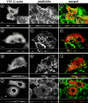
Mutated ezrin loses its cell membrane localization in transfected A431 and COS1 cells. Human adenocarcinoma A431 epithelioid cells (a–f) and monkey kidney COS1 fibroblasts (g–l) were transfected with either VSV-G–tagged wild-type ezrin (a–c and g–i) or mutated ezrin (K253N, K254N, K262N, and K263N) (d–f and j–l) DNA and treated for indirect Texas red localization of ezrin with anti–VSV antibody (left) and F-actin with FITC-coupled phalloidin (middle). Pictures represent the projected maximum intensities of several focal planes ranging from substrate to apical level, except for d and j. Vertical xz sections at the level of the dotted lines are under each figure. Insets are enlargements of the squared areas in a–c. Dual localizations of ezrin and actin are merged in color, colocalization appearing in yellow. Transfected wild-type ezrin is located in actin-rich cell surface structures such as microspikes, dorsal microvilli (a–c, circles) or ruffles (g–i, arrows), and in lateral cell membranes (a and c, intercellular contacts). Note that focal adhesion plaques (b, c, e, and f, insets, arrowheads) and stress fibers (k) are devoid of any ezrin. Transfected mutated ezrin is essentially located as a cytosolic network (d, f, j, and l; note that single focal planes are used only in d and j for clearer visualization of ezrin), but is also faintly detectable on the cell membranes in COS1 cells (j, arrow). Localization of wild-type ezrin in actin-rich dorsal structures and mutated ezrin in cytoplasm is evident in xz sections. Bars, 10 μm.
Figure 3
Figure 3
Cosedimentation of wild-type and mutant ezrin 1-333 with large liposomes containing different amounts of PIP2. Wild-type (WT) and mutant ezrin domains (55 μg protein/ml) were incubated in the presence of 100 mM NaCl and in the absence or presence of large liposomes containing 5, 10, or 20% of PIP2, the remainder being PC (0.5 mg lipid/ml). The fraction of cosedimenting protein was assessed as described in Materials and Methods. Mean ± SD of three to eight experiments.
Figure 4
Figure 4
The mutations introduced specifically target ezrin-PIP2 interactions. (A) Introduction of four K > N mutations in ezrin did not change the chymotrypsin digestion pattern. Wild-type (WT) and mutated ezrin 1-586 (K253N, K254N, K262N, and K263N) (285 μg/ml) were incubated in the presence or absence of 3.3 μg/ml chymotrypsin at room temperature for the indicated times. After SDS-PAGE, membranes were immunoblotted with a polyclonal antibody targeted against the first 310 amino acids of ezrin. No major difference in the digestion patterns was observed when wild-type or mutated ezrin were compared. (B) Introduction of four K > N mutations in ezrin (K253N, K254N, K262N, and K263N) did not alter F-actin binding. F-actin binding was measured using a solid phase assay as described by Roy et al. 1997. Actin concentrations were chosen so that they were below, around, and above the dissociation constant (500 nM) for actin binding to ezrin. For each actin concentration, binding was measured in the presence or absence of PIP2. Bound actin was detected using a monoclonal anti-actin antibody. The top two blots illustrate the binding of actin to the NH2-terminal domain of wild-type and mutant ezrin (residues 1–333). The bottom two blots correspond to actin binding to full-length ezrin with or without the four K > N mutations. No obvious difference in F-actin binding capacity was observed when comparing mutated ezrin constructs with their parent wild-type counterparts. (C) Mutagenesis of the PIP2 binding site in ezrin 1-333 did not impair the interaction of the NH2-terminal domain of ezrin with the cytoplasmic tail of EBP50. The binding of wild-type ezrin 1-333 (WT and *) or mutated ezrin 1-333 (mutated and **) (1 μM) to GST-EBP 50 or GST was assessed as described for binding to CD44 in Materials and Methods. (Top) Coomassie staining, (bottom) Western blotting. The experiment had been done in triplicate for each construct. Western blotting showed that there was no major difference between WT ezrin 1-333 and the mutated form concerning binding to GST-EBP50. The right two lanes demonstrate the lack of binding of ezrin 1-333 WT (*) or mutated (**) when assayed with the control GST free of fusion protein. (D) Introduction of up to six K > N mutations in ezrin did not affect homotypic interactions. Wild-type ezrin 1-586 was coated in wells of a microtiter plate (Roy et al. 1997). Ezrin 1-333 constructs (0.5 μM each) were then added in F-actin buffer, except for the first lane, where no construct was added (*). Similar amounts of ezrin 1-586 were coated in each well as judged by the Coomassie stain of the top blot. The same polyclonal anti-ezrin antibody as in A was used to detect the construct overlaid and, irrespective of the mutations introduced, no major difference in the amount of ezrin 1-333 construct bound to ezrin 1-586 was observed (bottom). When no ezrin 1-586 was coated, no ezrin 1-333 was detectable (not shown). (E) Intrinsic tryptophane fluorescence was not modified upon introduction of four K > N mutations in full-length ezrin. (Top curve) Full-length mutated ezrin K253N, K254N, K262N, and K263N; (bottom curve) wild-type ezrin 1-586. Note that neither the maximum intensity of tryptophane fluorescence nor the wavelength for maximum emission were significantly affected upon introduction of the four mutations.
Figure 5
Figure 5
Mutagenesis of the PIP2 binding site in ezrin results in a loss of PIP2 requirement of the interaction of ezrin with the cytoplasmic tail of CD44. Binding of full-length wild-type ezrin or ezrin K253N, K254N, K262N, and K263N (K253,254,262,263N) to GST or GST-CD44 in the absence or presence of PIP2 was assessed as described in Materials and Methods. Aliquots of the supernatant were then analyzed by SDS-PAGE and immunoblotted for the presence of ezrin. (A) Coomassie blue staining of the blots shows that comparable amounts of GST-CD44 (or GST) were detected in all lanes (top, incubation with GST-CD44 beads; bottom, incubation with GST beads). In contrast to wild-type (WT) ezrin (top left), K253N, K254N, K262N, and K263N (K253,254,262,263N) mutations in ezrin induced constitutive binding to GST-CD44–coupled agarose beads independently of the addition of PIP2 micelles (top right). GST beads alone (bottom) were used as control. During the GST-CD44 preparation, significant cleavage of the fusion protein occurred, resulting in large amount of GST adsorbed on the gel. (B) The amount of wild-type or mutated ezrin bound to GST-CD44 or to GST alone was quantified by densitometry of the immunoblots shown in A. Black and hatched columns represent ezrin bound to GST and GST-CD44, respectively. The results shown are representative of three independent experiments.
Figure 7
Figure 7
Subcellular localization of transfected mutated and wild-type ezrin in COS1 cells. Transfected cells were fractionated as described in Materials and Methods and the resulting fractions were analyzed by SDS-PAGE and immunoblotting. Before immunodecoration, blots were stained with Coomassie blue to ensure that protein loadings were comparable. (A) Transfection of full-length ezrin. Transfected VSV-tagged wild-type and K253N, K254N, K262N, and K263N (K253,254,262,263N) ezrin were recovered in cytosol (C), Triton X-100 extractable (E), or nonextractable (N) material, as revealed after immunoblotting with anti–VSV-G mAb. Anti-ezrin antibody, which recognizes both endogenous and VSV-G–tagged ezrin, was used as a control (top). *Internal control with recombinant wild-type ezrin (100 ng load). Results shown are representative of three independent experiments. (B) Transfection of ezrin 1-310. Detection of the top blot was performed with an antibody against the NH2-terminal portion of ezrin. The proportion of cytosolic full-length ezrin (#) detected with this antibody is lower than that presented in A due to the poor accessibility of the anti–N antibody to the epitopes in the full-length ezrin. Note that some degradation of ezrin was detectable in the nonextractable material (N). Immunodetection of the bottom blot was performed with anti–VSV-G mAb.
Figure 8
Figure 8
The mutated NH2-terminal domain of ezrin partly loses its interactions with membranes. Monkey COS1 fibroblasts were transfected with either VSV-G–tagged wild-type (a–c) or mutated (K253N, K254N, K262N, and K263N) (d–f) ezrin 1-310 and treated for immunolocalization of actin (b and e) and VSV (a and d) as in Fig. 6. Transfected wild-type NH2-terminal ezrin was mainly localized in actin-rich structures such as dorsal microvilli and ruffles (b and c, arrows; xz sections, circles). It induces formation of long filopodia containing both actin (b and c, arrowheads) and transfected ezrin (a and c, arrowheads). Note the presence of nodes along filopodia, particularly rich in NH2-terminal ezrin (open arrows). The transfected mutated form of NH2-terminal ezrin was predominantly localized in the cytoplasm (d and f), although some mutant ezrin localized in the same actin-rich structures (e and f) as wild-type NH2-terminal ezrin. Staining of nuclei was observed only in cells transfected with mutated ezrin 1-310 (d and f). Bars, 10 μm.
Figure 9
Figure 9
Location of the targeted lysines in moesin. The crystallographic data of moesin were treated with the molecular visualization program RASMOL (R. Sayle, Glaxo Wellcome, Greenford, UK). (A) The different domains of the molecule defined by Pearson et al. 2000 were colored as follows: residues 4–94, violet (domain FI); residues 95–201, blue (domain F2); residues 202–297, green (domain F3); and residues 488–577, red (COOH-terminal domain). The lysines of interest have been highlighted in yellow. The distances between the epsilon NH2 of lysines 63–262, 63–254, and 254–263 are equal to 38.1, 27.6, and 19 Å, respectively. Note that all these lysines are accessible to solvent and therefore to PIP2. They are all located on the same side of the molecule. (B) Moesin residues that may be in direct contact with the bilayer. Using RASMOL, residues exposed on the surface of moesin and enclosed by the triangle formed by lysines 63, 64, 253, 254, 262, and 263 (bold and underlined) have been identified (bold).
Figure 9
Figure 9
Location of the targeted lysines in moesin. The crystallographic data of moesin were treated with the molecular visualization program RASMOL (R. Sayle, Glaxo Wellcome, Greenford, UK). (A) The different domains of the molecule defined by Pearson et al. 2000 were colored as follows: residues 4–94, violet (domain FI); residues 95–201, blue (domain F2); residues 202–297, green (domain F3); and residues 488–577, red (COOH-terminal domain). The lysines of interest have been highlighted in yellow. The distances between the epsilon NH2 of lysines 63–262, 63–254, and 254–263 are equal to 38.1, 27.6, and 19 Å, respectively. Note that all these lysines are accessible to solvent and therefore to PIP2. They are all located on the same side of the molecule. (B) Moesin residues that may be in direct contact with the bilayer. Using RASMOL, residues exposed on the surface of moesin and enclosed by the triangle formed by lysines 63, 64, 253, 254, 262, and 263 (bold and underlined) have been identified (bold).

References

    1. Algrain M., Turunen O., Vaheri A., Louvard D., Arpin M. Ezrin contains cytoskeleton and membrane binding domains accounting for its proposed role as a membrane-cytoskeletal linker. J. Cell Biol. 1993;120:129–139. - PMC - PubMed
    1. Amieva M.R., Litman P., Huang L., Ichimaru E., Furthmayr H. Disruption of dynamic cell surface architecture of NIH3T3 fibroblasts by the N-terminal domains of moesin and ezrinin vivo imaging with GFP fusion proteins. J. Cell Sci. 1999;112:111–125. - PubMed
    1. Andréoli C., Martin M., Leborgne R., Reggio H., Mangeat P. Ezrin has properties to self-associate at the plasma membrane. J. Cell Sci. 1994;107:2509–2521. - PubMed
    1. Blomberg N., Nilges M. Functional diversity of PH domainsan exhaustive modelling study. Fold. Des. 1997;2:343–355. - PubMed
    1. Bradford M.M. A rapid and sensitive method for the quantitation of microgram quantities of protein utilizing the principle of protein-dye binding. Anal. Biochem. 1976;72:248–254. - PubMed

Publication types

MeSH terms