. 2000 Nov 27;151(5):1119-28.
doi: 10.1083/jcb.151.5.1119.

Phosphorylation of ADF/cofilin abolishes EGF-induced actin nucleation at the leading edge and subsequent lamellipod extension

Affiliations

Phosphorylation of ADF/cofilin abolishes EGF-induced actin nucleation at the leading edge and subsequent lamellipod extension

N Zebda et al. J Cell Biol. .

Abstract

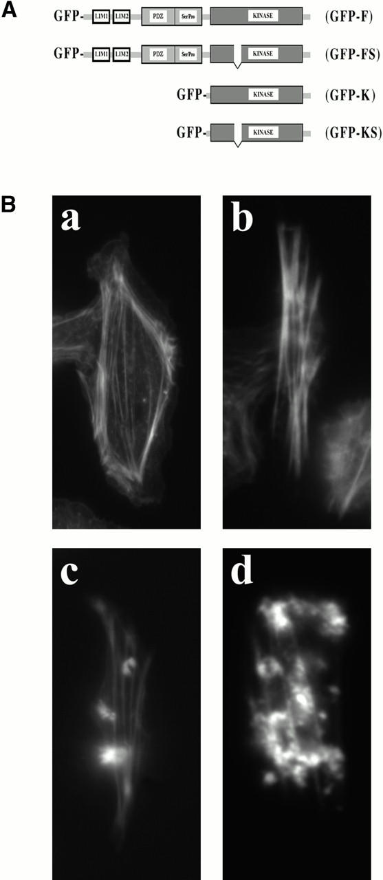
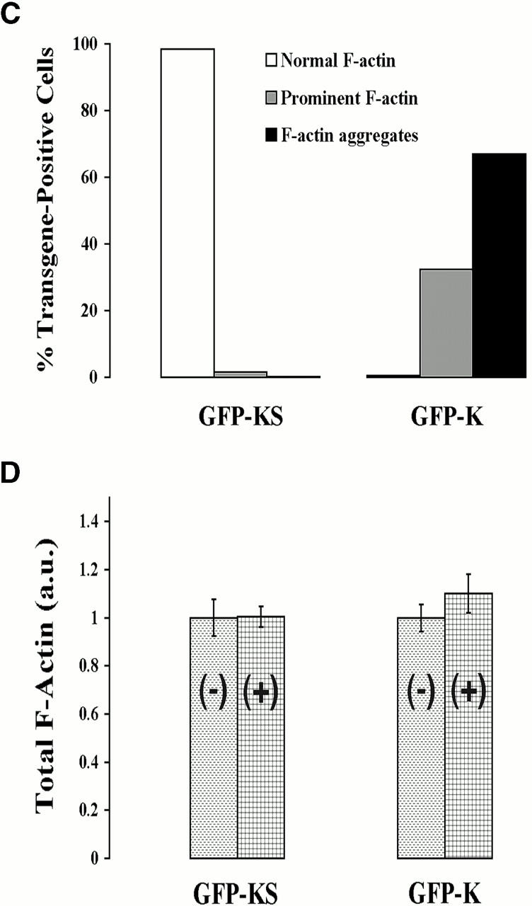
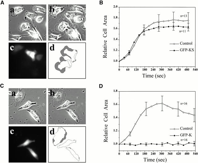
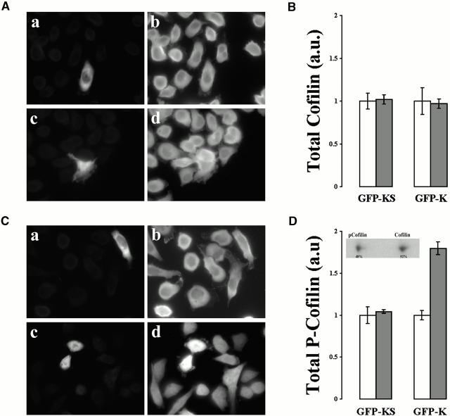
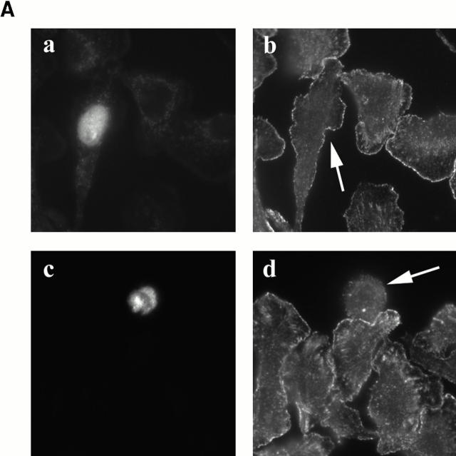
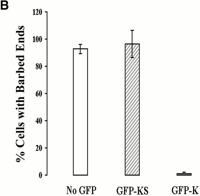
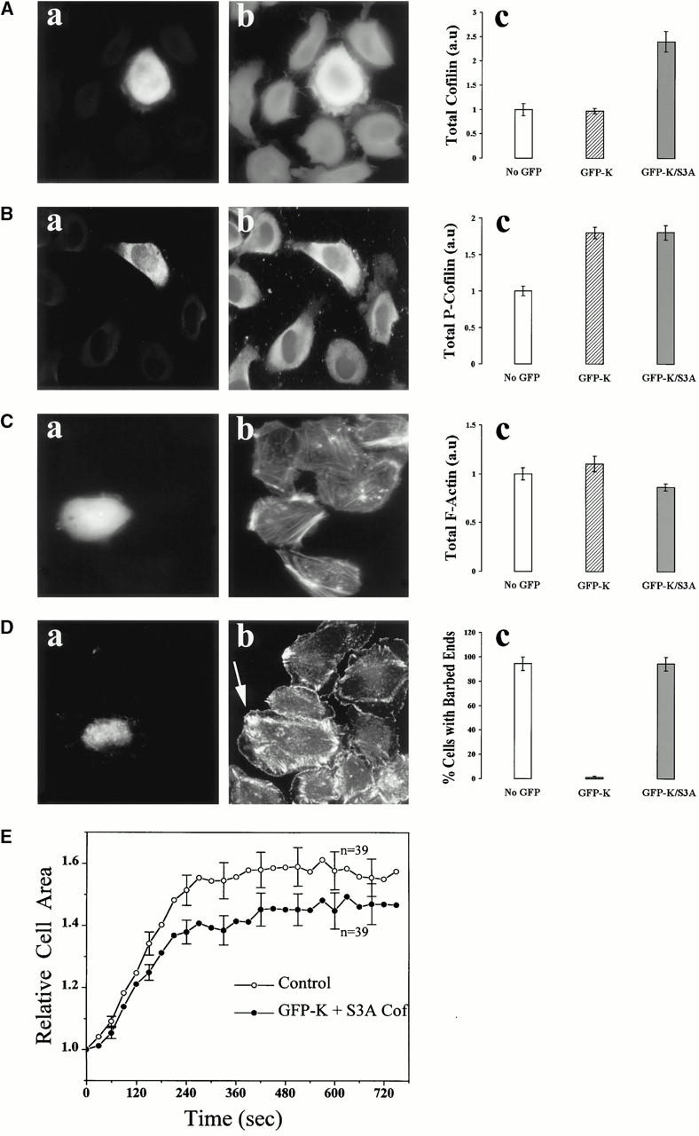
In metastatic rat mammary adenocarcinoma cells, cell motility can be induced by epidermal growth factor. One of the early events in this process is the massive generation of actin barbed ends, which elongate to form filaments immediately adjacent to the plasma membrane at the tip of the leading edge. As a result, the membrane moves outward and forms a protrusion. To test the involvement of ADF/cofilin in the stimulus-induced barbed end generation at the leading edge, we inhibited ADF/cofilin's activity in vivo by increasing its phosphorylation level using the kinase domain of LIM-kinase 1 (GFP-K). We report here that expression of GFP-K in rat cells results in the near total phosphorylation of ADF/cofilin, without changing either the G/F-actin ratio or signaling from the EGF receptor in vivo. Phosphorylation of ADF/cofilin is sufficient to completely inhibit the appearance of barbed ends and lamellipod protrusion, even in the continued presence of abundant G-actin. Coexpression of GFP-K, together with an active, nonphosphorylatable mutant of cofilin (S3A cofilin), rescues barbed end formation and lamellipod protrusion, indicating that the effects of kinase expression are caused by the phosphorylation of ADF/cofilin. These results indicate a direct role for ADF/cofilin in the generation of the barbed ends that are required for lamellipod extension in response to EGF stimulation.


Figures

Figure 1
Figure 1
Expression of the GFP-K induces aggregation of F-actin and formation of stress fibers without significantly increasing the F-actin content. (A) A schematic diagram is shown of the GFP-tagged constructs used in this study. (B) Cells were subdivided into three categories and scored accordingly: cells exhibiting similar phalloidin staining compared with (a, normal F-actin) transgene-negative cells; (b, prominent F-actin) cells with thicker stress fibers; and (c and d, F-actin aggregates) cells containing F-actin aggregates. (C) Quantitative analysis of the three categories in cells expressing the kinase domain of LIM kinase 1 (n = 176) or the corresponding inactive construct GFP-KS (n = 64). (D) Total F-actin was measured in phalloidin-stained cells expressing GFP-K (n = 118) and GFP-KS (n = 64). Fluorescence intensities are plotted as mean ± SEM here and in all figures. The data are representative for three experiments. The same results were obtained here and in all figures, regardless of the tag used (GFP or Myc), of the method used to deliver the DNA constructs (lipofection or microinjection), and whether the cDNA-microinjected cells were incubated in the absence of serum before fixation.
Figure 1
Figure 1
Expression of the GFP-K induces aggregation of F-actin and formation of stress fibers without significantly increasing the F-actin content. (A) A schematic diagram is shown of the GFP-tagged constructs used in this study. (B) Cells were subdivided into three categories and scored accordingly: cells exhibiting similar phalloidin staining compared with (a, normal F-actin) transgene-negative cells; (b, prominent F-actin) cells with thicker stress fibers; and (c and d, F-actin aggregates) cells containing F-actin aggregates. (C) Quantitative analysis of the three categories in cells expressing the kinase domain of LIM kinase 1 (n = 176) or the corresponding inactive construct GFP-KS (n = 64). (D) Total F-actin was measured in phalloidin-stained cells expressing GFP-K (n = 118) and GFP-KS (n = 64). Fluorescence intensities are plotted as mean ± SEM here and in all figures. The data are representative for three experiments. The same results were obtained here and in all figures, regardless of the tag used (GFP or Myc), of the method used to deliver the DNA constructs (lipofection or microinjection), and whether the cDNA-microinjected cells were incubated in the absence of serum before fixation.
Figure 4
Figure 4
Expression of the GFP-K abolishes EGF-induced lamellipod extension. MTLn3 cells were transiently transfected with either (A and B) GFP-KS or (C and D) GFP-K. 16–24 h after transfection, the cells were starved for 3 h and stimulated with 5 nM EGF. In A (GFP-KS) and C (GFP-K), live-phase contrast images were taken (a) before and (b) 4 min after EGF was added to the cells. Tracing of select cells from a and b were merged to show (d, grey area) the EGF-induced cell area increase, and (c) the GFP fluorescence is shown to indicate transgene-positive cells. In B (GFP-KS) and D (GFP-K), continuous time lapse digital images were analyzed using DIAS software. The cell area is a measure of lamellipod extension.
Figure 2
Figure 2
Expression of GFP-K phosphorylates almost all cofilin in vivo without changing cofilin expression levels. MTLn3 cells microinjected with expression vectors for (a and b) GFP-KS or (c and d) GFP-K were immunostained with antibodies to (A) cofilin or to (C) phosphocofilin/ADF. Cofilin and phosphocofilin/ADF staining are shown in b and d, and transgene-positive cells are visualized in a and c. (B and D) Fluorescence intensities (mean ± SEM) for GFP-positive cells (solid bars) were normalized against neighboring transgene-negative cells (open bars). The data presented are representative for at least three experiments. The inset in D is presented as a reference showing the relative amounts of cofilin (52%) versus phosphocofilin (48%) in MTLn3 cells.
Figure 3
Figure 3
Expression of the GFP-K abolishes EGF-induced actin barbed end generation at the leading edge. (A) MTLn3 cells were microinjected with either (a and b) GFP-KS or (c and d) GFP-K cDNAs and assayed for EGF-induced actin nucleation activity. Incorporation of exogenous-labeled actin in the cell cortex is seen as a thin rim of fluorescence at the cell periphery. (A, a and c, and arrows in A, b and d) Cells expressing exogenous kinase were detected by direct visualization of the GFP green fluorescence. Due to saponin-permeabilization before fixation, the cytoplasmic GFP-kinase is extracted, leaving behind GFP-kinase that is mostly nuclear and perinuclear in location. (B) The percent of cells showing incorporation of exogenous actin at the leading edge was determined for GFP-K cells (n = 96), GFP-KS cells (n = 28), and transgene-negative cells. The data presented is representative of at least three experiments.
Figure 3
Figure 3
Expression of the GFP-K abolishes EGF-induced actin barbed end generation at the leading edge. (A) MTLn3 cells were microinjected with either (a and b) GFP-KS or (c and d) GFP-K cDNAs and assayed for EGF-induced actin nucleation activity. Incorporation of exogenous-labeled actin in the cell cortex is seen as a thin rim of fluorescence at the cell periphery. (A, a and c, and arrows in A, b and d) Cells expressing exogenous kinase were detected by direct visualization of the GFP green fluorescence. Due to saponin-permeabilization before fixation, the cytoplasmic GFP-kinase is extracted, leaving behind GFP-kinase that is mostly nuclear and perinuclear in location. (B) The percent of cells showing incorporation of exogenous actin at the leading edge was determined for GFP-K cells (n = 96), GFP-KS cells (n = 28), and transgene-negative cells. The data presented is representative of at least three experiments.
Figure 5
Figure 5
Expression of the S3A mutant of cofilin rescues the inhibition of barbed ends and lamellipod extension caused by the kinase domain. MTLn3 cells were microinjected either with GFP-K or a 1:1 mixture of GFP-K and S3A cofilin cDNAs and analyzed 5–8 h later. (A–C) Cells were immediately fixed and processed for staining with (A, b) anti-cofilin antibody Ab287, (B, b) anti-phosphocofilin/ADF antibody, or (C, b) bodipy-phalloidin. (D and E) Cells were starved for 3 h before adding EGF. (D) Barbed end generation was measured as described in the legend to Fig. 3. (E) Lamellipod extension was scored as described in the legend to Fig. 4. (A–D, a, and D, b, arrow) Expressing cells are identified by direct visualization of the GFP fluorescence. Quantitative analyses are shown in A–D, c. “No-GFP” are control transgene-negative cells. The number of GFP-K/S3A cofilin–expressing cells (solid bars) analyzed in A–D (c) is 27, 63, 78, and 26, respectively. The data presented are representative of at least three experiments.

Similar articles

Cited by

References

    1. Aizawa H., Sutoh K., Yahara I. Overexpression of cofilin stimulates bundling of actin filaments, membrane ruffling, and cell movement in Dictyostelium . J. Cell Biol. 1996;132:335–344. - PMC - PubMed
    1. Arber S., Brbayannis F.A., Hanser H., Schneider C., Stanyon C.A., Bernard O., Caroni P. Regulation of actin dynamics through phosphorylation of cofilin by LIM-kinase 1. Nature. 1998;393:805–809. - PubMed
    1. Bailly M., Yan L., Whitesides G.M., Condeelis J.S., Segall J.E. Regulation of protrusion shape and adhesion to the substratum during chemotactic responses of mammalian carcinoma cells. Exp. Cell Res. 1998;241:285–299. - PubMed
    1. Bailly M., Macaluso F., Cammer M., Chan A., Segall J.E., Condeelis J.S. Relationship between Arp2/3 complex and the barbed ends of actin filaments at the leading edge of carcinoma cells after epidermal growth factor stimulation. J. Cell Biol. 1999;145:331–345. - PMC - PubMed
    1. Bamburg J.R. Proteins of the ADF/Cofilin familyessential regulators of actin Dynamics. Annu. Rev. Cell. Dev. Biol. 1999;15:185–230. - PubMed

Publication types

MeSH terms