. 2000 Nov;106(10):1229-37.
doi: 10.1172/JCI11066.

Estrogen deficiency induces bone loss by enhancing T-cell production of TNF-alpha

Affiliations

Estrogen deficiency induces bone loss by enhancing T-cell production of TNF-alpha

S Cenci et al. J Clin Invest. 2000 Nov.

Abstract

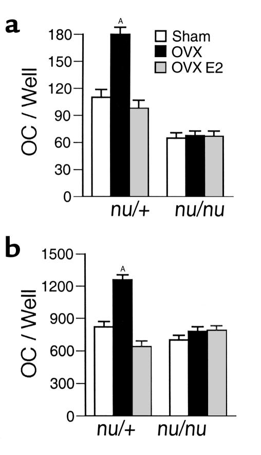
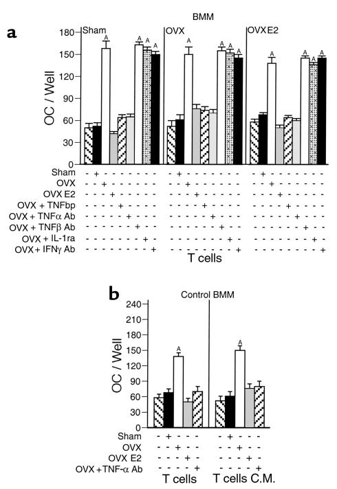
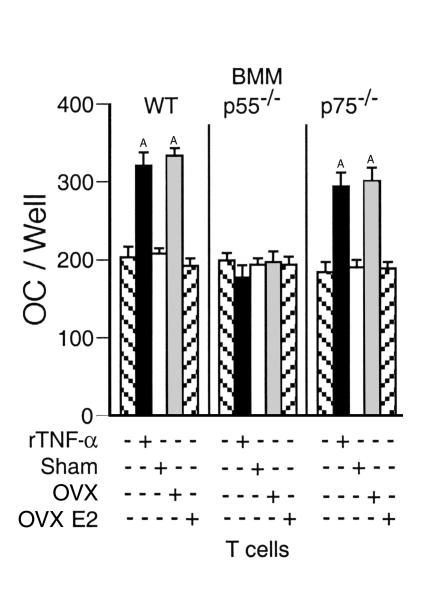
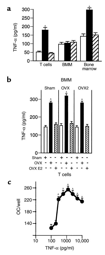
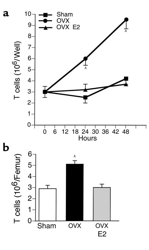
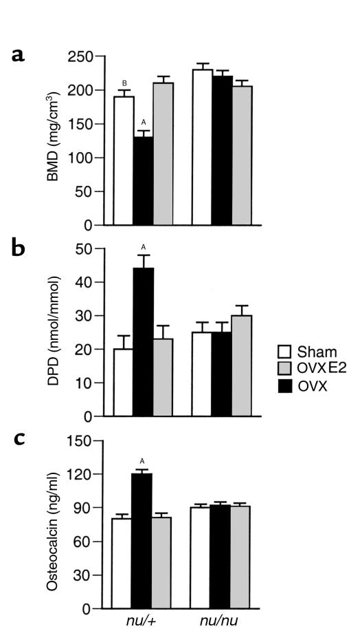
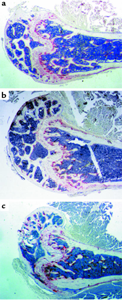
Estrogen deficiency induces bone loss by upregulating osteoclastogenesis by mechanisms not completely defined. We found that ovariectomy-enhanced T-cell production of TNF-alpha, which, acting through the TNF-alpha receptor p55, augments macrophage colony-stimulating factor-induced (M-CSF-induced) and RANKL-induced osteoclastogenesis. Ovariectomy failed to induce bone loss, stimulate bone resorption, or increase M-CSF- and RANKL-dependent osteoclastogenesis in T-cell deficient mice, establishing T cells as essential mediators of the bone-wasting effects of estrogen deficiency in vivo. These findings demonstrate that the ability of estrogen to target T cells, suppressing their production of TNF-alpha, is a key mechanism by which estrogen prevents osteoclastic bone resorption and bone loss.


Figures

Figure 1
Figure 1
Ovariectomy fails to increase osteoclast formation in cultures of bone marrow cells from nude mice. Mean (± SEM) number of osteoclasts from three independent experiments (n = 6 wells per group). AP < 0.05 compared to estrogen replete mice. (a) Unfractionated bone marrow cells (106 cells/well) from OVX and estrogen-replete nude and euthymic mice were harvested 4 weeks after surgery and cultured for 7 days with 1,25(OH)2D3 (10–8M) to induce osteoclast formation. (b) Bone marrow cells were depleted of stromal cells by adherence for 12 hours. Nonadherent bone marrow cells (106 cells/well) were cultured for 7 days with optimal osteoclastogenic concentrations of M-CSF (25 ng/ml) and RANKL (100 ng/ml). OC, osteoclast.
Figure 2
Figure 2
T cells from OVX mice produce a soluble factor upregulating RANKL- and M-CSF–induced osteoclast formation, which is blocked by TNF-α neutralization. Mean (± SEM) of three independent experiments (n = 6 wells per group). AP < 0.05 compared with all other groups. (a) Cocultures of purified monocytes and purified T cells from OVX and estrogen-replete mice stimulated with optimal osteoclastogenic concentrations of M-CSF and RANKL. No osteoclast formation was observed in RANKL- and M-CSF–stimulated cultures of T cells in the absence of BMMs. Similarly, osteoclast differentiation of BMMs was not induced by stimulation with either RANKL or M-CSF alone, both in the presence or the absence of T cells (not shown). (b) Purified monocytes from intact mice stimulated with optimal osteoclastogenic concentrations of M-CSF and RANKL were cultured with either purified T cells from OVX and estrogen-replete mice or T cell–conditioned media (C.M.) from OVX and estrogen-replete mice. AP < 0.05 compared with all other groups.
Figure 3
Figure 3
T cells from OVX mice fail to increase RANKL- and M-CSF–induced osteoclast differentiation of monocytes lacking the p55 TNF-α receptor. Mean (± SEM) number of osteoclasts from three independent experiments (n = 6 wells per group). BMMs were purified from C57BL/6 mice lacking either TNF-α receptor 1 (p55–/–) or TNF-α receptor II (p75–/–) and WT controls of the same genetic background and cocultured with T cells derived from OVX or estrogen-replete WT mice in the presence of optimal concentrations of RANKL and M-CSF. AP < 0.05 compared with all groups.
Figure 4
Figure 4
Ovariectomy increases T-cell secretion of TNF-α. (a) TNF-α levels secreted by purified BMMs and T cells. Mean (± SEM) levels in culture supernatants from three independent experiments (n = 6 wells per group). P < 0.05 compared with all other groups. Culture media was collected after 3 days and TNF-α measured by ELISA as described (15). (b) TNF-α levels in cocultures of BMMs and T cells. Mean (± SEM) levels in culture supernatants from three independent experiments (n = 6 wells per group). P < 0.05 compared with all other groups. (c) TNF-α augments RANKL- and M-CSF–induced osteoclast formation in a dose-dependent manner. Mean (± SEM) number of osteoclasts from three independent experiments (n = 6 wells per dose). Purified monocytes from control mice were stimulated with M-CSF (25 ng/ml), RANKL (100 ng/ml), and TNF-α (0–10 ng/ml), and cultured for 7 days. AP < 0.05 compared with control.
Figure 5
Figure 5
Ovariectomy increases T-cell proliferation and bone marrow T-cell content. Mean (± SEM) of three independent experiments (n = 6 wells per group). AP < 0.05 compared with baseline. (a) Unstimulated T cells purified from OVX and estrogen-replete mice were cultured for 48 hours. At the end of the culture, T cells were counted. (b) Freshly isolated whole bone marrow was analyzed by FACS using anti-CD90 Ab. Total number of bone marrow cells was counted. Results are expressed as number of T cells per femur.
Figure 6
Figure 6
Ovariectomy fails to induce bone loss and upregulate bone turnover in nude mice (n = 6 per group). AP < 0.05 compared with all other groups. BP < 0.05 compared with sham nu/nu. (a) Trabecular BMD (mean ± SEM) of the tibia was measured 4 weeks after surgery in excised tibiae using pQCT. (b) DPD excretion (mean ± SEM) at 4 weeks after surgery. (c) Serum osteocalcin levels (mean ± SEM) at 4 weeks after surgery.
Figure 7
Figure 7
Histology of nude mice femur. Note that sham (top panel), OVX (middle panel), and estrogen-treated OVX (bottom panel) had similar cortical thickness and lack of trabecular bone resorption below the growth plates. The three groups had similar number of TRAP-positive osteoclasts (stained in red).

Comment in

References

    1. Lacey DL, et al. Osteoprotegerin ligand is a cytokine that regulates osteoclast differentiation and activation. Cell. 1998;93:165–176. - PubMed
    1. Kong YY, et al. OPGL is a key regulator of osteoclastogenesis, lymphocyte development and lymph-node organogenesis. Nature. 1999;397:315–323. - PubMed
    1. Yasuda H, et al. Osteoclast differentiation factor is a ligand for osteoprotegerin/osteoclastogenesis-inhibitory factor and is identical to TRANCE/RANKL. Proc Natl Acad Sci USA. 1998;95:3597–3602. - PMC - PubMed
    1. Manolagas SC, Jilka RL. Bone marrow, cytokines, and bone remodeling. N Engl J Med. 1995;332:305–311. - PubMed
    1. Pacifici R. Estrogen, cytokines and pathogenesis of postmenopausal osteoporosis. J Bone Miner Res. 1996;11:1043–1051. - PubMed

Publication types

MeSH terms

Substances