. 2000 Nov;106(10):1271-80.
doi: 10.1172/JCI10793.

Protective effects of anti-C5a in sepsis-induced thymocyte apoptosis

Affiliations

Protective effects of anti-C5a in sepsis-induced thymocyte apoptosis

R F Guo et al. J Clin Invest. 2000 Nov.

Abstract

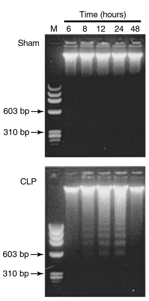
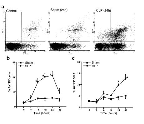
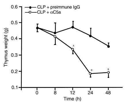
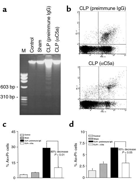
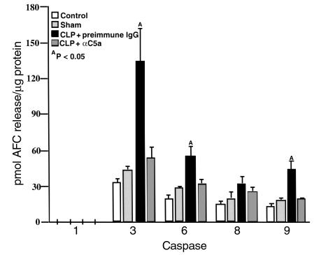
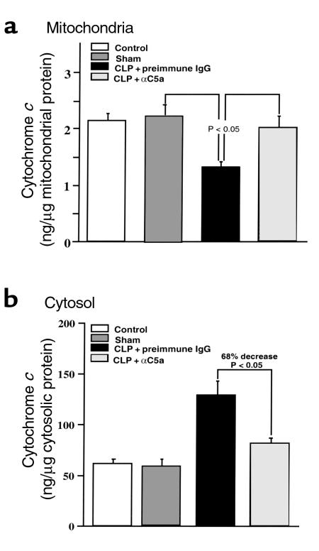
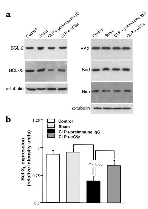
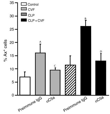
Multiorgan apoptosis occurs during sepsis. Following cecal ligation and puncture (CLP) in rats, thymocytes underwent apoptosis in a time-dependent manner. C5a blockade dramatically reduced thymocyte apoptosis as measured by thymic weight, binding of annexin V to thymocytes, and laddering of thymocyte DNA. When C5a was generated in vivo by infusion of purified cobra venom factor (CVF), thymocyte apoptosis was significantly increased. Similar results were found when CVF was injected in vivo during the early stages of CLP. In animals 12 hours after induction of CLP, there was an increase in the activities of caspase-3, -6, and -9, but not caspase-1 and -8. Cytosolic cytochrome c levels increased by twofold, whereas mitochondrial levels showed a 50% decrease. Western blot analysis revealed that the content of Bcl-X(L) (but not of Bcl-2, BAX, Bad, and Bim) significantly decreased in thymocytes after CLP. C5a blockade in the sepsis model almost completely inhibited caspase-3, -6, and -9 activation, significantly preserved cytochrome c in the mitochondrial fraction, and restored Bcl-X(L) expression. These data suggest that systemic activation of complement induces C5a-dependent apoptosis of thymocytes and that the blockade of C5a during sepsis rescues thymocytes from apoptosis.


Figures

Figure 1
Figure 1
Time course of CLP-induced DNA fragmentation in thymocytes. DNA was extracted from thymocytes of sham or CLP rats at indicated hours and was subjected to agarose gel electrophoresis. M, DNA molecular weight marker. The results are representative of three separate experiments.
Figure 2
Figure 2
Flow cytometric analysis of Ax (FITC→) and PI (PI→) staining in thymocytes from sham or CLP rats. Thymocytes isolated from sham or CLP rats at the times indicated were stained with Ax-FITC and PI. (a) FACS analysis of apoptotic thymocytes from normal, sham, and CLP rats 24 hours after surgery. The regions for Ax-positive only (Ax+PI, lower right), PI-positive only (AxPI+, upper left), double-positive (Ax+PI+, upper right), and double-negative (AxPI, lower left) were established as described in Methods. (b) The percentage of early-stage apoptotic thymocytes during the course of 48 hours after sham or CLP-induced sepsis. (c) The percentage of late apoptotic and early necrotic thymocytes over 48 hours after sham or CLP-induced sepsis. AP < 0.05 when compared with the sham group. BP < 0.01 when compared with the sham group. Values represent means ± SEM (n = 3 or 4 animals).
Figure 3
Figure 3
The effects of C5a blockade on thymus atrophy induced by CLP. Immediately after CLP surgery, animals were treated intravenously with 400 μg preimmune IgG or 400 μg anti-rat C5a IgG (αC5a). At the indicated time points, the animals were sacrificed and the thymus was weighed. The same protocol for C5a blockade was used in all subsequent figures. For each vertical bar, n = 4 or 5 animals. AP < 0.05 when compared with IgG control. BP < 0.01 when compared to IgG control.
Figure 4
Figure 4
Protection of thymocytes from apoptosis by C5a blockade in CLP-induced sepsis. Twenty-four hours after surgery, thymocytes were harvested from control, sham, CLP-treated with preimmune IgG, and anti-C5α–treated CLP rats, and evaluated for DNA fragmentation and Ax/PI staining using the same protocols described for Figures 1 and 2. (a) Effect of C5a blockade on DNA fragmentation induced by CLP, representative of three separate experiments. (b) Quantitative analysis of the effect of C5a blockade on thymocyte apoptosis by flow cytometry. The percentages of early-stage apoptotic cells (c) and late apoptotic/early necrotic cells (d) are presented as means ± SEM (n = 4 animals).
Figure 5
Figure 5
Inhibition of caspase activation in thymocytes by in vivo C5a blockade in CLP-induced sepsis. Cell lysates were incubated with caspase substrates: Ac-YVAD-AFC (caspase-1), Ac-DEVD-AFC (caspase-3), Ac-VEID-AFC (caspase-6), Ac-IETD-AFC (caspase-8), and Ac-LEHD-AFC (caspase-9). The cleaved AFC was detected on a Cytofluor II plate reader. The results were presented by picomoles of AFC released from substrates per microgram protein over a 2-hour incubation time. AP < 0.05 when compared with sham or CLP + αC5a group. All values represent means ± SEM (n = 4).
Figure 6
Figure 6
Effects of C5a blockade on cytochrome c release from mitochondria in thymocytes during CLP-induced sepsis. Cytosolic and mitochondrial fractions were separated as described in methods. (a and b) Cytochrome c concentrations in cytosol and mitochondria were quantitatively determined by ELISA. P < 0.05 when compared with sham or CLP + αC5a group. All values represent means ± SEM (n = 4).
Figure 7
Figure 7
Effects of C5a blockade on expression of Bcl-2 family members in thymocytes during CLP-induced sepsis. (a) The cytoplasmic extracts containing 100 μg protein were electrophoresized in a denaturing polyacrylamide gel and then transferred to a nitrocellulose membrane. Protein expression was evaluated by Western blot using the following antibodies: polyclonal rabbit anti-Bcl-2, polyclonal rabbit anti-BAX, monoclonal mouse anti-Bcl-XL, polyclonal rabbit anti-Bad, polyclonal rabbit anti-Bim, and monoclonal mouse anti-α-tubulin. The graphs are representative of three separate experiments. (b) Densitometric quantification of Bcl-XL expression normalized to α-tubulin. All values represent means ± SEM (n = 3). P < 0.05 when compared with sham or CLP + αC5a group.
Figure 8
Figure 8
Effects of C5a blockade on NF-κB activation in thymocytes from CLP animals. (a) Supershift analysis of CLP-induced NF-κB components. DNA-binding reactions with nuclear extracts from thymocytes harvested from rats 12 hours after CLP were incubated with 32P-labeled NF-κB oligonucleotide in the absence or presence of antibodies to the NF-κB proteins indicated (p50, p52, p65, p68, p75). Supershift of p65 is indicated by a filled arrowhead. (b) Activation of NF-κB in sepsis and effect of C5a blockade. NF-κB activation was evaluated in thymocytes harvested from rats in various groups as indicated. Each lane represents a different animal. The filled arrow indicates NF-κB p65. The open arrow indicates NF-κB p50. The open arrowhead indicates an unknown band.
Figure 9
Figure 9
Effects of C5a blockade on thymocyte apoptosis induced by CVF. Rats were injected intravenously with 3 U CVF and 500 μg preimmune IgG or 500 μg anti-C5a, and thymocyte apoptosis was evaluated by Ax staining 3 hours after infusion. For CLP rats, CVF was given 3 hours after CLP surgery. AP < 0.05 when compared with control or CVF + αC5a group. BP < 0.01 when compared with CLP or CLP + CVF + αC5a group. All values represent means ± SEM (n = 4 or 5). CP > 0.05 when compared with control. DP > 0.05 when compared with CLP.

References

    1. Darville T, Giroir B, Jacobs R. The systemic inflammatory response syndrome (SIRS): immunology and potential immunotherapy. Infection. 1993;21:279–290. - PubMed
    1. Papathanassoglou ED, Moynihan JA, Ackerman MH. Does programmed cell death (apoptosis) play a role in the development of multiple organ dysfunction in critically ill patients? A review and a theoretical framework. Crit Care Med. 2000;28:537–549. - PubMed
    1. Ayala A, Chaudry IH. Immune dysfunction in murine polymicrobial sepsis: mediators, macrophages, lymphocytes and apoptosis. Shock. 1996;6:S27–S38. - PubMed
    1. Mahidhara R, Billiar TR. Apoptosis in sepsis. Crit Care Med. 2000;28:N105–N113. - PubMed
    1. Hotchkiss RS, et al. Apoptotic cell death in patients with sepsis, shock, and multiple organ dysfunction. Crit Care Med. 1999;27:1230–1251. - PubMed

Publication types

MeSH terms

Substances