. 2000 Nov;106(10):1281-90.
doi: 10.1172/JCI7236.

Altered podocyte structure in GLEPP1 (Ptpro)-deficient mice associated with hypertension and low glomerular filtration rate

Affiliations

Altered podocyte structure in GLEPP1 (Ptpro)-deficient mice associated with hypertension and low glomerular filtration rate

B L Wharram et al. J Clin Invest. 2000 Nov.

Abstract

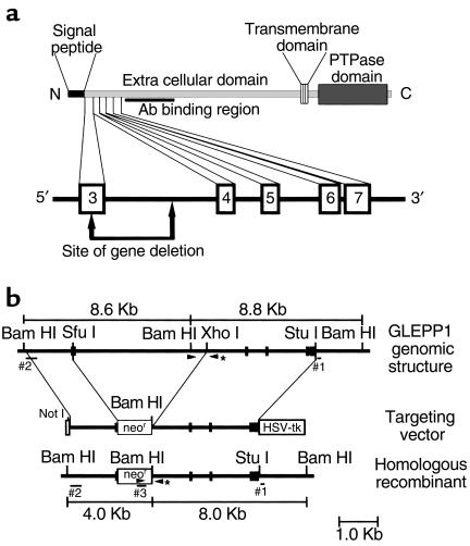
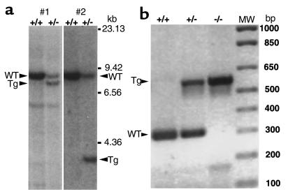
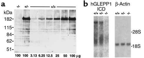
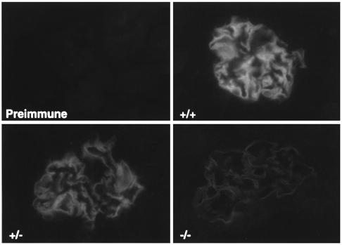
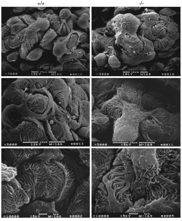
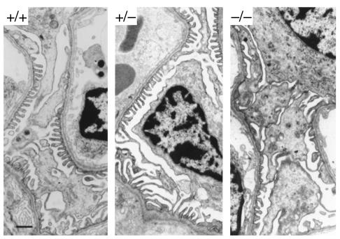
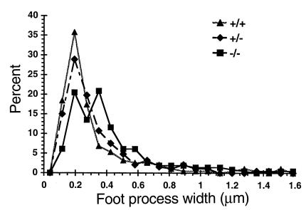
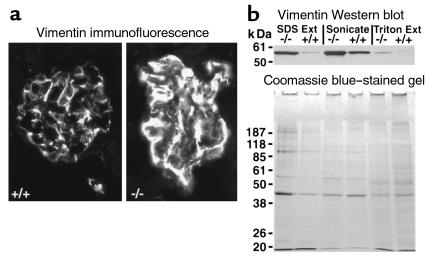
Glomerular epithelial protein 1 (GLEPP1) is a receptor tyrosine phosphatase present on the apical cell surface of the glomerular podocyte. The GLEPP1 gene (PTPRO:) was disrupted at an exon coding for the NH(2)-terminal region by gene targeting in embryonic stem cells. Heterozygote mating produced the expected genotypic ratio of 1:2:1, indicating that the Ptpro(-/-) genotype does not lead to embryonic or neonatal lethality. Kidney and glomerular structure was normal at the gross and light microscopic levels. Scanning and transmission electron microscopy showed that Ptpro(-/-) mice had an amoeboid rather than the typical octopoid structure seen in the wild-type mouse podocyte and that there were blunting and widening of the minor (foot) processes in association with altered distribution of the podocyte intermediate cytoskeletal protein vimentin. Reduced filtration surface area in association with these structural changes was confirmed by finding reduced glomerular nephrin content and reduced glomerular filtration rate in Ptpro(-/-) mice. There was no detectable increase in the urine albumin excretion of Ptpro(-/-) mice. After removal of one or more kidneys, Ptpro(-/-) mice had higher blood pressure than did their wild-type littermates. These data support the conclusion that the GLEPP1 (Ptpro) receptor plays a role in regulating the glomerular pressure/filtration rate relationship through an effect on podocyte structure and function.


Figures

Figure 1
Figure 1
(a) Structure of 5′ end of the mouse GLEPP1 gene showing the exon/intron arrangement in relation to the coding region for the NH2-terminal end of the GLEPP1 protein. Numbered boxes represent exons. Exon 3 is a putative designation pending sequencing of the 5′ region of the gene. The site of gene deletion so as to interrupt an exon, induce a frameshift, and introduce a stop codon is shown. The Ab-binding site, which is 3′ of the deletion, is also shown. (b) Diagrammatic illustrations of the region of the wild-type allele to be targeted (upper), the GLEPP1 gene-targeting vector (middle), and the predicted structure of the targeted allele (lower). Filled boxes correspond to exons 3–7 depicted in a. Sites for restriction enzymes used for constructing the targeting vector and Southern screening of ES cell clones are shown. Size and location of Southern probes are shown, including the 3′ external probe (#1), the 5′ internal probe (#2), and the neo probe (#3). Arrowheads denote sites for PCR primers used for genotype analysis of mice. The 3′ primer, denoted by an asterisk, is common to both the wild-type and targeted alleles.
Figure 2
Figure 2
(a) Southern blots of BamHI-digested DNA showing lanes with wild-type (WT) and targeted (Tg) alleles hybridized with probes for the 3′ (#1) and 5′ (#2) ends of the targeted region as defined in Figure 1. (b) PCR analyses of tail DNA from Ptpro+/+, Ptpro+/–, and Ptpro–/– mice, showing the assay system used to identify genotypes.
Figure 3
Figure 3
(a) Western blot of proteins extracted from glomeruli isolated from Ptpro–/–, Ptpro+/–, and Ptpro+/+ mice. The GLEPP1 band is at 180 kDa. Bands at lower molecular weights are nonspecific and were seen at the same intensities on a control blot developed with preimmune Ab’s (not shown). Numbers below lanes correspond to micrograms of glomerular Triton protein extract loaded onto each lane. Ptpro+/+ extract was loaded at twofold serial dilutions from the right side of the gel and could be detected at the 32-fold dilution. GLEPP1 was not detectable in Ptpro–/– extracts. GLEPP1 was present in Ptpro+/– extracts at a level approximately one-half that of extracts from Ptpro+/+ mice. (b) Northern blot of RNA prepared from glomeruli isolated from Ptpro+/+, Ptpro+/–, and Ptpro–/– mice. Left panel: probed with 1 kb human GLEPP1 intracellular domain, random-primed DNA labeled with α-32P dCTP. Right panel: probed with human β-actin probe to show equal loading of RNA onto the lanes. No detectable hybridization was seen in the Ptpro–/– RNA sample.
Figure 4
Figure 4
Indirect immunofluorescence of mouse renal cortex cryostat sections developed using rabbit anti-rat GLEPP1 ECD IgG (Ptpro+/+, upper right; Ptpro+/–, lower left; and Ptpro–/–, lower right). Preimmune IgG tested on a Ptpro+/+ renal cortical section is shown at upper left. The photomicrographs are developed to the same intensity of signal. ×400.
Figure 5
Figure 5
Scanning electron micrographs of glomeruli made at different magnifications, with the wild-type (Ptpro+/+) mice shown in the left three panels and Ptpro–/– mice shown in the right three panels. The major processes seen in Ptpro–/– mice were broader, wider, and less distinct than the narrower, more well-defined major processes seen in the wild-type mice. The overall structure of the Ptpro–/– podocytes is more amoeba-like compared with the octopus-like structure of the wild-type (×3,000, top panels). At high power the foot processes of the Ptpro+/+ mice can be seen to be regular and fingerlike in appearance in contrast to the broader, shorter (more toelike) foot processes found in the Ptpro–/– mice (×10,000, bottom panels).
Figure 6
Figure 6
Transmission electron micrographs showing glomerular capillary loops from Ptpro+/+, Ptpro+/–, and Ptpro–/– mice, revealing broader foot process structure in the Ptpro–/– mice. Bar, 1 μm.
Figure 7
Figure 7
Graph of the distribution of foot-process width measured on TEM in regions of open glomerular capillaries from Ptpro+/+ (triangle), Ptpro+/– (diamond), and Ptpro–/– (square) animals. The Ptpro+/+ and Ptpro+/– mice have narrow foot processes predominantly in the 0.2-μm range, whereas the Ptpro–/– mice have a significantly broader range of foot-process width.
Figure 8
Figure 8
(a) Immunofluorescence photomicrographs developed with anti-vimentin Ab’s in Ptpro+/+ (left) and Ptpro–/– (right) glomeruli showing the different distribution of vimentin and apparent increase in amount in the Ptpro–/– mice. (b) Western blot of glomerular extracts from Ptpro–/– and Ptpro+/+ mice made using differential extraction by Triton alone (right two lanes), Triton plus sonication to solubilize cytoskeletal proteins (middle two lanes), and the SDS-extracted pellet (left two lanes). The signal for vimentin (top) suggests that vimentin is present in increased amount in the Ptpro–/– mice. Approximately equal protein loading of glomerular extracts is shown by Coomassie blue–stained gels (bottom).

References

    1. Drenckhahn D, Franke RP. Ultrastructural organization of contractile and cytoskeletal proteins in glomerular podocytes of chicken, rat and man. Lab Invest. 1988;59:673–682. - PubMed
    1. Reiser J, Kriz W, Kretzler M, Mundel P. The glomerular slit diaphragm is a modified adherens junction. J Am Soc Nephrol. 2000;11:1–8. - PubMed
    1. Kerjaschki D, Sharkey DJ, Farquhar MG. Identification and characterization of podocalyxin, the major sialoprotein of the renal glomerular epithelial cell. J Cell Biol. 1984;98:1591–1596. - PMC - PubMed
    1. Gelberg H, Healy L, Whiteley H, Miller LA, Vimr E. In vivo enzymatic removal of the α2→6-linked sialic acid from the glomerular filtration barrier results in podocyte charge alteration and glomerular injury. Lab Invest. 1996;74:907–920. - PubMed
    1. Kreidberg JA, et al. Alpha 3 beta 1 integrin has a crucial role in kidney and lung organogenesis. Development. 1996;122:3537–3547. - PubMed

Publication types

MeSH terms