Comparative Study
. 2000 Dec;59(12):945-53.
doi: 10.1136/ard.59.12.945.

Comparative study of the synovial histology in rheumatoid arthritis, spondyloarthropathy, and osteoarthritis: influence of disease duration and activity

Affiliations
Comparative Study

Comparative study of the synovial histology in rheumatoid arthritis, spondyloarthropathy, and osteoarthritis: influence of disease duration and activity

D Baeten et al. Ann Rheum Dis. 2000 Dec.

Abstract

Objectives: To compare the macroscopic and microscopic characteristics of synovial tissue in rheumatoid arthritis (RA), spondyloarthropathy (SpA), and osteoarthritis (OA) after exclusion of possible biases induced by disease duration or activity, or both.

Methods: Synovial biopsy specimens were obtained by needle arthroscopy in patients with early RA (n=16), late RA (n=14), early SpA (n=23), and OA (n=12). Macroscopic and microscopic features were scored on a four point scale and analysed as a function of disease duration (early versus late RA), local and systemic disease activity, and diagnosis.

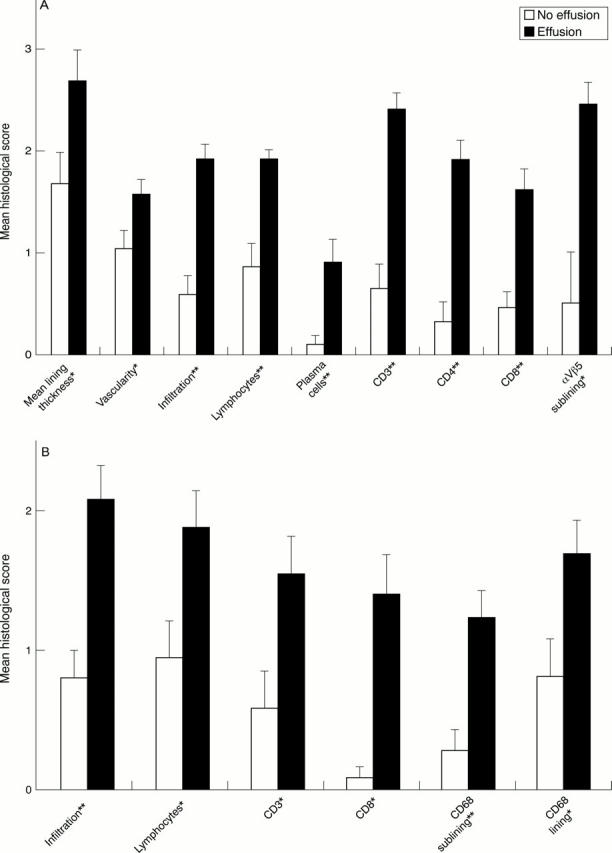
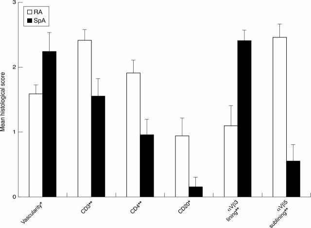
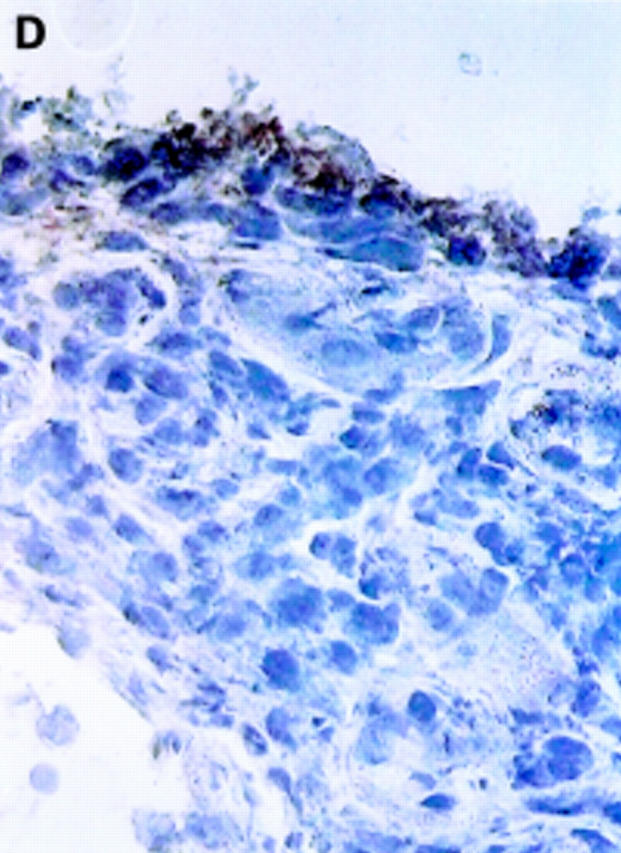
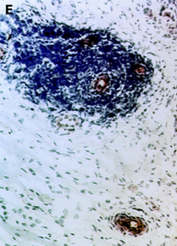
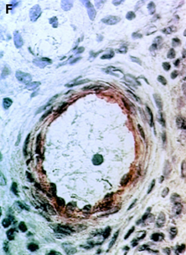
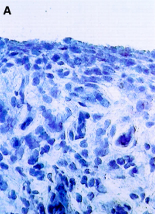
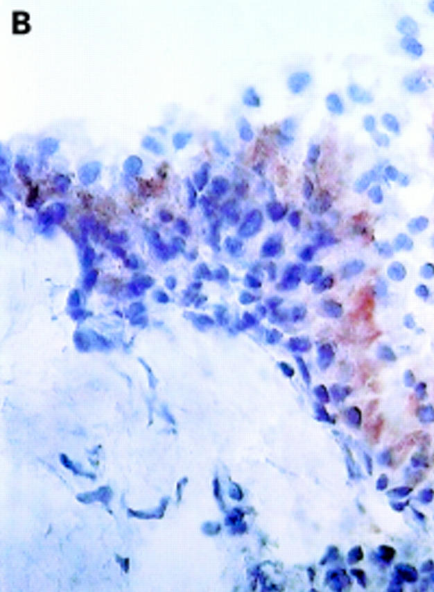
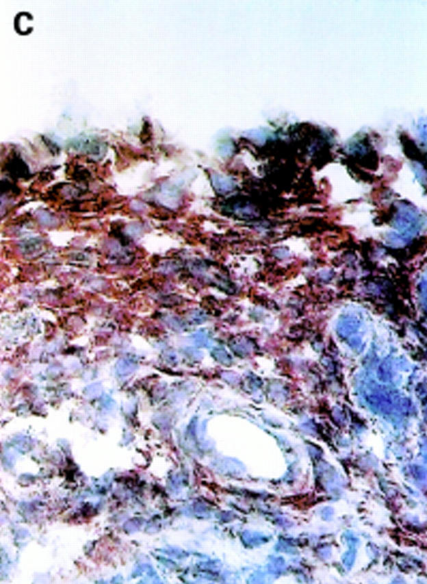
Results: Except for the maximal synovial lining thickness, no significant differences were seen between early and late RA. For disease activity, synovial histology was only weakly correlated with C reactive protein in RA, but seemed to be strongly dependent on effusion of the biopsied joint in all disease groups. After stratification for local disease activity, no disease related differences were found in patients without joint effusion. In contrast, important differences were found between patients with RA and SpA with active joint effusion. Synovial vascularity was macroscopically increased in SpA versus RA (p=0.017). A straight vessel pattern was only seen in RA, while tortuous vessels were preferentially seen in SpA. Vascularity was also microscopically increased in SpA compared with RA (p=0.031), and correlated with the macroscopic vascularity (r(s)=0.36, p=0.036). CD3+ (p=0.008), CD4+ (p=0.008), and CD20+ (p=0.024) lymphocytes were overrepresented in RA compared with SpA. The integrin expression in RA was characterised by a decrease of alphaVbeta3 in the synovial lining (p=0.006) and an increase of alphaVbeta5 in the sublining (p<0.001).

Conclusions: The immune architecture of the synovial membrane is more dependent on local disease activity than on disease duration. Synovium obtained from clinically affected joints shows important histological differences between RA and SpA.


Figures

Figure 1
Figure 1
Comparison of the histological scores in patients with and without effusion of the biopsied joint. Mean (standard error of the mean). *p<0.05. **p<0.01. (A) Patients with rheumatoid arthritis with (n=18) and without joint effusion (n=12). (B) Patients with spondyloarthropathy with (n=13) and without joint effusion (n=10).
Figure 2
Figure 2
Comparison of the histological scores in 18 patients with rheumatoid arthritis (RA) and 13 patients with spondyloarthropathy (SpA) with effusion of the biopsied joint. Mean (standard error of the mean). *p<0.05. **p<0.01.
Figure 3
Figure 3
αV integrin expression in the synovial membrane. Frozen sections of synovial biopsy specimens from RA (A and C) and SpA (B and D) were stained immunohistochemically for αVβ3 (A and B) and αVβ5 (C and D). (A) αVβ3 expression in RA synovium: no staining of synovial lining cells. (B) αVβ3 expression in SpA synovium: staining of synovial lining cells. (C) αVβ5 expression in RA synovium: staining of superficial synovial lining cells and of sublining cells. (D) αVβ5 expression in SpA synovium: staining of superficial synovial lining cells, but not of sublining cells. (E) αVβ3 expression on endothelial cells in RA synovium. (F) αVβ3 expression on endothelial cells in SpA synovium.
Figure 3
Figure 3
αV integrin expression in the synovial membrane. Frozen sections of synovial biopsy specimens from RA (A and C) and SpA (B and D) were stained immunohistochemically for αVβ3 (A and B) and αVβ5 (C and D). (A) αVβ3 expression in RA synovium: no staining of synovial lining cells. (B) αVβ3 expression in SpA synovium: staining of synovial lining cells. (C) αVβ5 expression in RA synovium: staining of superficial synovial lining cells and of sublining cells. (D) αVβ5 expression in SpA synovium: staining of superficial synovial lining cells, but not of sublining cells. (E) αVβ3 expression on endothelial cells in RA synovium. (F) αVβ3 expression on endothelial cells in SpA synovium.

Comment in

References

    1. Clin Rheumatol. 1999;18(6):434-41 - PubMed
    1. Scand J Immunol. 1992 Feb;35(2):159-65 - PubMed
    1. Arthritis Rheum. 2000 Jan;43(1):38-47 - PubMed
    1. Arthritis Rheum. 2000 Feb;43(2):259-69 - PubMed
    1. Clin Exp Rheumatol. 2000 Mar-Apr;18(2):201-7 - PubMed

Publication types

MeSH terms