. 2000 Dec;59(12):959-65.
doi: 10.1136/ard.59.12.959.

Apoptosis in normal and osteoarthritic human articular cartilage

Affiliations

Apoptosis in normal and osteoarthritic human articular cartilage

F Héraud et al. Ann Rheum Dis. 2000 Dec.

Abstract

Objectives: To investigate whether apoptosis occurs in osteoarthritis (OA), and if this phenomenon is modulated by human recombinant interleukin 1beta (hrIL1beta).

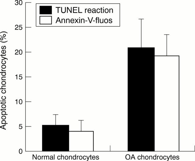
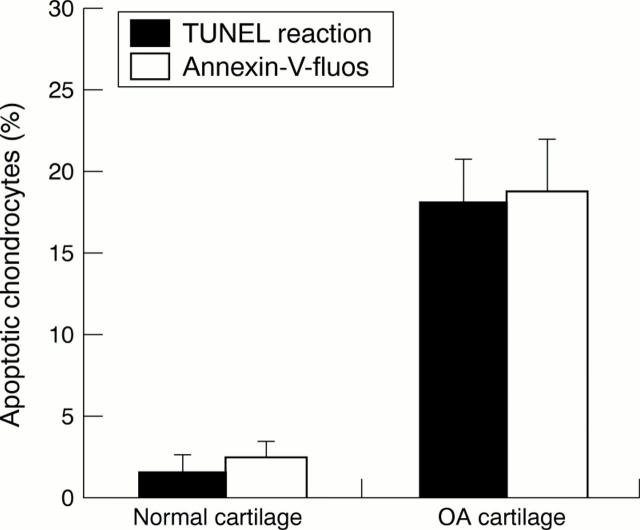
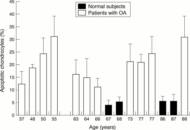
Methods: Human articular cartilage samples were obtained at the time of hip arthroplasty because of femoral neck fracture (normal cartilage) (n=4) or advanced coxarthrosis (OA cartilage) (n=14). Apoptotic chondrocytes, isolated by collagenase digestion and cultivated for 24 hours, or present in situ in frozen cartilage sections, were quantified by fluorescent microscopy using two apoptosis markers: the TUNEL reaction, which detects nuclear DNA fragmentation, and Annexin-V-fluos, which labels at the membrane level the externalisation of phosphatidylserine.

Results: In OA cartilage 18-21% of chondrocytes showed apoptotic features, compared with 2-5% in normal cartilage. The results were similar for the two comparative studies (in situ and in vitro) and for both apoptosis markers. Moreover, hrIL1beta increased the apoptosis rate in vitro in a dose dependent manner in OA and normal chondrocytes.

Conclusion: These results suggest that apoptosis may be an important factor in the evolution of OA and may be a new target for treatment of OA.


Figures

Figure 1
Figure 1
Detection of apoptosis by immunofluorescence on isolated OA chondrocytes by collagenase digestion (A and C) and in situ on fresh frozen OA cartilage (B and D). (A and B) Chondrocytes were fixed, permeabilised, and processed by the TUNEL reaction mixture. (C and D) Samples were treated with Annexin-V-fluos for 15 minutes and analysed immediately. An example of an apoptotic cell is identified by the arrows. (Original magnification A, B, C ×200; D ×100).
Figure 2
Figure 2
Comparative study of normal and OA chondrocytes using the TUNEL reaction and Annexin-V labelling in chondrocytes isolated by collagenase digestion. The ratio of apoptotic cells/total cells was calculated as a percentage. Values are means (SEM) from four experiments for normal chondrocytes and 11 experiments for OA chondrocytes (p<0.01).
Figure 3
Figure 3
Comparative study of normal and OA cartilage using the TUNEL reaction and Annexin-V labelling on frozen cartilage sections. The ratio of apoptotic cells/total cells was calculated as a percentage. Data are representative results from four experiments with normal cartilage and 13 experiments with OA cartilage (p<0.01).
Figure 4
Figure 4
Induction of apoptosis in OA chondrocytes by human recombinant interleukin 1β (hrIL1β). (A) Untreated control OA human chondrocytes. (B) OA human articular chondrocytes in primary culture stimulated with 10 ng/ml of hrIL1β for 24 hours and processed with Annexin-V-fluos for 15 minutes. Arrows identify examples of apoptotic cells. (Original magnification ×200.)
Figure 5
Figure 5
Percentage of apoptotic chondrocytes as a function of aging in the patients with OA and normal patients. Cells were isolated by collagenase digestion, and apoptosis in these isolated cells was analysed by TUNEL reaction labelling. Values are means (SEM) for 11 patients with OA and four normal patients.

References

    1. Anat Rec. 1998 Apr;250(4):418-25 - PubMed
    1. Am J Epidemiol. 1998 Mar 15;147(6):516-22 - PubMed
    1. Arthritis Rheum. 1998 Aug;41(8):1343-55 - PubMed
    1. Arthritis Rheum. 1998 Sep;41(9):1632-8 - PubMed
    1. J Histochem Cytochem. 1999 Feb;47(2):209-20 - PubMed