. 2000 Dec 5;97(25):13943-8.
doi: 10.1073/pnas.220389697.

Molecular determinants of the functional interaction between syntaxin and N-type Ca2+ channel gating

Affiliations

Molecular determinants of the functional interaction between syntaxin and N-type Ca2+ channel gating

I Bezprozvanny et al. Proc Natl Acad Sci U S A. .

Abstract

Syntaxin is a key presynaptic protein that binds to N- and P/Q-type Ca(2+) channels in biochemical studies and affects gating of these Ca(2+) channels in expression systems and in synaptosomes. The present study was aimed at understanding the molecular basis of syntaxin modulation of N-type channel gating. Mutagenesis of either syntaxin 1A or the pore-forming alpha(1B) subunit of N-type Ca(2+) channels was combined with functional assays of N-type channel gating in a Xenopus oocyte coexpression system and in biochemical binding experiments in vitro. Our analysis showed that the transmembrane region of syntaxin and a short region within the H3 helical cytoplasmic domain of syntaxin, containing residues Ala-240 and Val-244, appeared critical for the channel modulation but not for biochemical association with the "synprint site" in the II/III loop of alpha(1B). These results suggest that syntaxin and the alpha(1B) subunit engage in two kinds of interactions: an anchoring interaction via the II/III loop synprint site and a modulatory interaction via another site located elsewhere in the channel sequence. The segment of syntaxin H3 found to be involved in the modulatory interaction would lie hidden within the four-helix structure of the SNARE complex, supporting the hypothesis that syntaxin's ability to regulate N-type Ca(2+) channels would be enabled after SNARE complex disassembly after synaptic vesicle exocytosis.


Figures
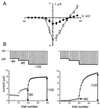
Figure 1
Figure 1
Coexpression of syntaxin 1A with N-type Ca2+ channels in Xenopus oocytes inhibited channel activity. (A) Peak current–voltage relationships of N-type Ca2+ channels (α1Bβ3α2) expressed in Xenopus oocytes, in isolation (open circles, n = 4) or with coexpressed syntaxin 1A (filled circles, n = 4). Symbols and error bars display mean ± SEM. (B) N-type Ca2+ channel inactivation properties analyzed by use of a “descending staircase” stimulation protocol. Comparison between behavior of channels expressed in isolation (Left) and in the presence of syntaxin 1A (Right). Inward Ba2+ currents were evoked by 50-ms depolarizing pulses to 0 mV from a holding potential of −60 mV. After 10 test pulses, holding potential was changed to −80 mV for 10 more test pulses, then further changed to −120 mV for 20 additional test pulses. The interpulse interval (10 seconds) and the test pulse level (0 mV) were kept constant throughout the experiment.
Figure 2
Figure 2
The carboxyl-terminal TMR of syntaxin 1A is essential for N-type Ca2+ channel modulation. The mutant constructs shown (Left) are described in Results and Materials and Methods. Striped areas denote various TMRs taken from syntaxin 2, syntaxin 2′, syntaxin 3, and the carboxyl-terminal region of syntaxin 2′′. Crosshatched area denotes a scrambled version of the TMR of s1A. Bar graph displays mean ± SEM values of the I80/I120 ratio for each construct (number of oocytes shown in parentheses).
Figure 3
Figure 3
The helical H3 domain of syntaxin 1A contains structural determinants critical for the modulatory interaction with N-type Ca2+ channels. (A) Bar graph displays mean ± SEM values of the I80/I120 ratio for each mutant construct (number of oocytes shown in parentheses). The mutant constructs are described in Materials and Methods. Domain deletions in s1A-C, M267, V248 M, s1A-M215, and s1A-I195 are as indicated. The small vertical lines denote point mutations in s1A-H3 point mutants. Of the various point mutant constructs, only s1A-YK6 shows an I80/I120 ratio significantly greater than wild-type s1A. For further details, see text. (B) In vivo 35S-Met labeling of s1A constructs expressed in Xenopus oocytes and immunoprecipitation with HPC-1 antibody. (C) Levels of 35S-Met labeling, normalized by expression level of wild-type syntaxin 1A in each experiment, shown as mean ± SEM.
Figure 4
Figure 4
Effect of deletions from the II/III loop of α1B influence N-type channel inactivation properties and affect responsiveness of N-type channels to syntaxin 1A modulation. (A) Voltage-dependent availability of wild-type N-type channels (filled circles, V1/2 = −76 mV), Nd2 (open squares, V1/2 = −68 mV), Nd5 (open circles, V1/2 = −36 mV), Nd6 (downward triangles, V1/2 = −35 mV), Nd7 (upward triangles, V1/2 = −41 mV). N-type channel mutant constructs are described in Materials and Methods. Data points are averages derived from at least three independent experiments for each channel mutant (error bars indicate ± SEM). (B) Analysis of midpoint shifts (ΔV1/2) produced by coexpression of syntaxin. (Left) Wild-type N-type channels (with synprint site shown as thick line), shown schematically above a series of II/III loop deletion constructs Nd2, Nd5, Nd6, Nd7 (extent of deletion indicated by thin dotted line). (Right) Coexpression with syntaxin (filled symbols) induced a hyperpolarizing shift in the inactivation properties of wild-type channels and each of the II/III loop mutants relative to their behavior in control (open symbols). The shift was estimated as the displacement along the voltage axis that would be needed to align a data point in the presence of syntaxin (left vertical arrow) with the voltage-dependent curve describing inactivation in the absence of syntaxin (right vertical arrow). Estimates of the shift were as follows: WT, ΔV1/2 > 28 mV; Nd2, ΔV1/2 =16 mV; Nd5, ΔV1/2 =12 mV; Nd6, ΔV1/2 =10 mV; Nd7, ΔV1/2 =12 mV. The shift was statistically significant (P < 0.01) in all cases. Where not shown, error bars (SEM) were smaller than the symbols.
Figure 5
Figure 5
Wild-type syntaxin 1A and s1AYK6 point mutant display similar affinity for the II/III loop of the N-type Ca2+ channel in a scintillation proximity displacement assay. 35S-labeled s1A-M267X binding to an immobilized recombinant GST fusion protein incorporating the II/III loop of α1B. Binding assays were performed in the presence of increasing concentrations of soluble competing proteins: wild-type syntaxin 1A fragment (s1A-M267X, square), a corresponding fragment of mutant s1A-YK6 (triangle), and a control protein (GST, circle). All three proteins were derived from bacterial expression and purified. In all scintillation proximity binding assays, nonspecific binding (determined by binding of 35S-s1A-M267X to immobilized GST) was about 23% of total binding (determined by maximal binding of 35S-s1A-M267X to immobilized GST-II/III loop fragment). The data points show mean ± SEM values for percent binding, expressed as a fraction of maximal binding (n = 3).

References

    1. Bennett M K, Calakos N, Scheller R H. Science. 1992;257:255–259. - PubMed
    1. Yoshida A, Oho C, Omori A, Kuwahara R, Ito T, Takahashi M. J Biol Chem. 1992;267:24925–24928. - PubMed
    1. Sollner T, Bennett M K, Whiteheart S W, Scheller R H, Rothman J E. Cell. 1993;75:409–418. - PubMed
    1. Sutton R B, Fasshauer D, Jahn R, Brunger A T. Nature (London) 1998;395:347–353. - PubMed
    1. Kee Y, Lin R C, Hsu S C, Scheller R H. Neuron. 1995;14:991–998. - PubMed

Publication types

LinkOut - more resources