. 2000 Dec 5;97(25):13755-9.
doi: 10.1073/pnas.240398797.

3' deletions cause aniridia by preventing PAX6 gene expression

Affiliations

3' deletions cause aniridia by preventing PAX6 gene expression

J D Lauderdale et al. Proc Natl Acad Sci U S A. .

Abstract

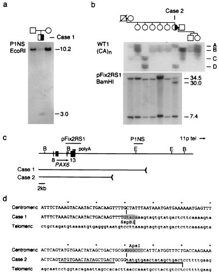
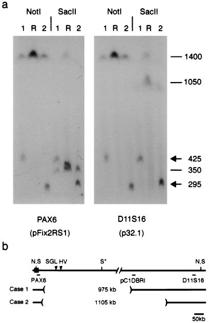
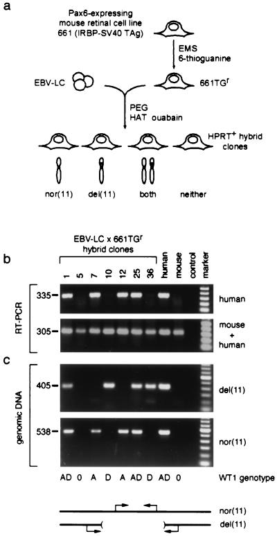
Aniridia is a panocular human eye malformation caused by heterozygous null mutations within PAX6, a paired-box transcription factor, or cytogenetic deletions of chromosome 11p13 that encompass PAX6. Chromosomal rearrangements also have been described that disrupt 11p13 but spare the PAX6 transcription unit in two families with aniridia. These presumably cause a loss of gene expression, by removing positive cis regulatory elements or juxtaposing negative DNA sequences. We report two submicroscopic de novo deletions of 11p13 that cause aniridia but are located >11 kb from the 3' end of PAX6. The clinical manifestations are indistinguishable from cases with chain-terminating mutations in the coding region. Using human x mouse retinoblastoma somatic cell hybrids, we show that PAX6 is transcribed only from the normal allele but not from the deleted chromosome 11 homolog. Our findings suggest that remote 3' regulatory elements are required for initiation of PAX6 expression.


Figures

Figure 1
Figure 1
Identification of 3′ PAX6 rearrangements in sporadic aniridia. (a) Southern analysis of case 1. Probe P1NS detects a novel 3.0-kb EcoRI fragment in the affected child, but not in his parents, and a 10.2-kb EcoRI fragment in normal DNA. (b) Haplotype and Southern analysis of case 2. (Upper) Segregation of the WT1 (CA)n repeat polymorphism. Four alleles are distinguished. Each allele is represented by an ensemble of fragments that differ from the mean length by 1–2 repeat units; these arise by replication slippage during PCR amplification. Case 2 is heterozygous (AD) at WT1. (Lower) Probe pFix2RS1 detects 34.5-kb and 7.4-kb BamHI fragments in normal DNA, and an additional 30-kb BamHI fragment was in the affected female, who is heterozygous. The mutation arose on a paternal chromosome 11 and is linked to WT1 allele D. (c) Restriction map showing PAX6 exons 8–13, polyadenylation sites (arrowheads), hybridization probes, and the centromeric deletion breakpoints in these two cases. The breakpoints are located 22.1 kb and 11.6 kb from the 3′ end of PAX6. E, EcoRI; B, BamHI. Some of the EcoRI sites have been omitted. (d) DNA sequence spanning the centromeric (uppercase) and telomeric (lowercase) breakpoints of cases 1 and 2 in normal DNA and the deletion junctions. The junction points are indicated by vertical lines and the 20-bp duplication in case 2 is underlined. The rearrangement in case 1 creates an SspBI restriction site at the junction.
Figure 2
Figure 2
Physical mapping of 3′ PAX6 deletions. (a) Pulsed-field gel of lymphoblastoid genomic DNA from cases 1 and 2 and a normal reference individual (R) hybridized with probes pFix2RS1 and p32.1. These probes detect one 1400-kb NotI (N) fragment and two SacII (S) fragments, which are 350 kb (centromeric) and 1050 kb (telomeric). The internal SacII (S*) site is partially cleaved (22, 32). The second SacII fragment in the reference (R) lane reflects a likely sequence polymorphism. (b) Restriction map showing hybridization probes and deletion breakpoints relative to PAX6. Probe pC1PBRI detects an altered NotI fragment in case 1 but not case 2 (data not shown). The deletions encompass breakpoints of the HV reciprocal translocation and the SGL paracentric inversion (arrowheads), which are located, respectively, 124 kb and 85–100 kb from the 3′ end of PAX6 (22, 33, 34).
Figure 3
Figure 3
Human PAX6 is not expressed from the deleted chromosome 11 in somatic cell hybrids. (a) Strategy used to make hybrid cell lines. EBV-lymphoblastoid cells from aniridia case 2 (EBV-LC) were fused to 661TGr, an HPRT-deficient mouse retinoblastoma cell line. Hybrid clones (n = 37) were selected in HAT + ouabain and classified on the basis of human chromosome 11 content. In the absence of selective pressure for human autosomes, both homologs were free to segregate. (b) RNA analysis of representative hybrids. Human PAX6 and mouse Pax6 expression is demonstrated by parallel RT-PCRs, using conditions that amplify human transcripts only or transcripts from both species (see Materials and Methods). Hybrid clones 1, 7, 12, and 25 retain the nor(11) and express PAX6, whereas clones 10 and 36 retain only the del(11) and do not express PAX6. Similar results were obtained for all 37 clones. Reactions performed on RNA from Ad12 (human) and 661TGr (mouse) retinal cells, and a no-RT control, are shown for comparison. The size of each PCR product is indicated in base pairs. (c) DNA analysis of representative hybrids. The chromosome 11 content is demonstrated by diagnostic genomic PCRs. An amplicon within the deletion identifies nor(11), and an amplicon spanning the breakpoint junction identifies del(11). PCRs performed on genomic DNA from case 2 (human) and 661TGr (mouse) cells, and a no-template control reaction, are shown for comparison. WT1 genotype data for the hybrid cell lines are indicated below the panels; the D allele is linked to the deletion. marker, 100-bp ladder (Roche); nor(11), normal; del(11), deleted.

References

    1. Gehring W J, Ikeo K. Trends Genet. 1999;15:371–377. - PubMed
    1. Ton C C, Hirvonen H, Miwa H, Weil M M, Monaghan P, Jordan T, van Heyningen V, Hastie N D, Meijers-Heijboer H, Drechsler M, et al. Cell. 1991;67:1059–1074. - PubMed
    1. Hanson I, Jordan T, Van Heyningen V. In: Molecular Genetics of Inherited Eye Disorders. Wright A F, Jay B, editors. Switzerland: Harwood Academic; 1994. pp. 445–467.
    1. Prosser J, van Heyningen V. Hum Mutat. 1998;11:93–108. - PubMed
    1. Glaser T, Walton D S, Maas R L. Nat Genet. 1992;2:232–239. - PubMed

Publication types

Associated data